Youtube

Advancing the Science of Ultrasound Guided Regional Anesthesia and Pain Medicine

Supraclavicular Block

Anatomy

The primary ventral rami of C5 and C6 unite to form the upper trunk above the subclavian artery, C7 becomes the middle trunk and C8 and T1 unite to form the lower trunk. Both the brachial plexus and the subclavian artery lie on top of the first rib. The brachial plexus is located lateral and posterior to the subclavian artery. The subclavian vein and anterior scalene muscle are found medial to the subclavian artery. The pleura is usually found within 1-2 cm from the brachial plexus.

The supraclavicular approach to the brachial plexus at the level of the nerve trunks or divisions was first described by Kulenkampf. However, the original technique was associated with a high incidence of pneumothorax. Although subsequent modification of this technique has lowered the complication rate, performance of this block without visual guidance is generally not recommended for outpatients.

1 = anterior scalene muscle
2 = middle scalene muscle
CL = clavicle
FR = first rib
SA = subclavian artery
SV = subclavian vein

Scanning Technique

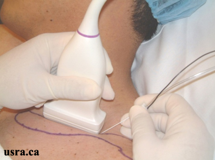
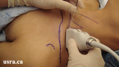
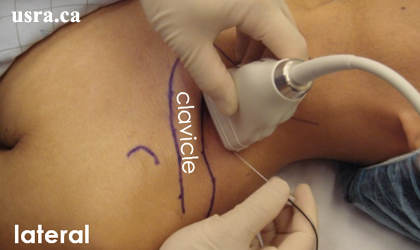
  • After skin and transducer preparation (see transducer preparation section), place a linear 38-mm, high frequency 10-15 MHz transducer firmly over the supraclavicular fossa in the coronal oblique plane to obtain the best possible transverse view of the subclavian artery and brachial plexus.
  • Position the patient supine with the head slightly turned to the contralateral side.
Transducer over left supraclavicular fossa
  • Optimize machine imaging capability by selecting the appropriate depth of field (within 2-3 cm), focus range and gain.
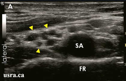
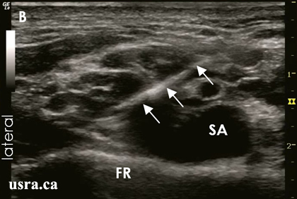
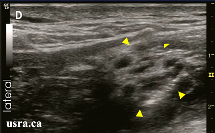
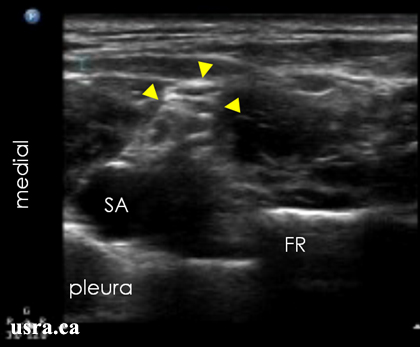
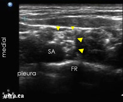
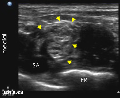
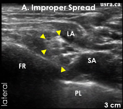
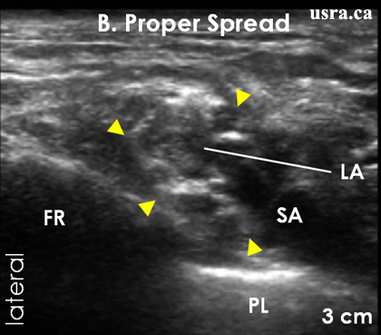
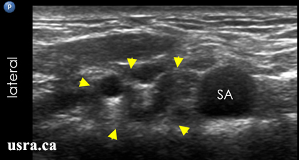
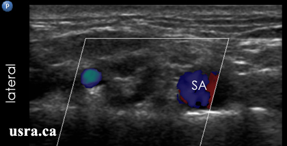
  • Visualize the trunks or divisions in the transverse view (short axis). Nerves in the supraclavicular region appear hypoechoic and are round or oval. The brachial plexus is located lateral and posterior to the pulsatile subclavian artery and superior to the first rib.

Anatomical Correlation

Arrowheads = trunks/divisions of the brachial plexus
C = clavicle
FR = first rib
SA = subclavian artery
SAM and SMM = scalenus anterior and medius muscles
SV = subclavian vein
White box = scanned area

Nerve Localization

  • Perform a systematic anatomical survey from medial to lateral and superficial to deep.
  • The brachial plexus (trunks) is generally easy to locate in this region. The subclavian artery serves as an easily identifiable reference point to locate the brachial plexus.
  • First locate the subclavian artery.
  • The subclavian vein is found more medially.
  • The anterior scalene muscle inserts onto the first rib between these 2 vessels.
  • Identify the hyperechoic first rib lying deep to the vessels and its bony shadow.
  • Identify the pleura and compare it with the hyperechoic first rib. Note air artifact, the “comet tail” sign and pleura sliding movement during respiration.
  • Note the skin-to-first rib and skin-to-pleura distance.
  • The brachial plexus is consistently found lateral and posterior to the subclavian artery and above the first rib.

Needle Insertion Approach

  • Ultrasound guided supraclavicular block is considered an INTERMEDIATE skill level block because real time observation of needle tip location during needle advancement is critical.
  • The In Plane (IP) approach is strongly recommended for this block. It is important to track the needle tip in real time to avoid inadvertent pleural puncture.

In Plane Approach (Lateral to Medial)

  • For the IP approach, insert a 5 cm 22G insulated block needle on the outer (lateral) end of the ultrasound transducer after skin local anesthetic infiltration. Advance the needle along the long axis of the transducer in the same plane as the ultrasound beam. In this way, the needle shaft and tip can be visualized in real time as the needle is advanced towards the target nerves.
The transducer is placed over the right supraclavicular fossa. The needle is inserted in plane with the ultrasound transducer and beam in a lateral to medial direction.
  • Confirm the identity of the nerves by electrical stimulation if desired. Useful stimulation endpoints for surgery proximal to the elbow are biceps and triceps twitches but aim to get hand muscle twitches for surgery distal to the elbow.
Arrows = block needle
Arrowheads = nerves
SA = subclavian artery
  • Note that this procedure is unique and is dramatically different from conventional supraclavicular techniques. With one hand holding the transducer and the other holding the needle, the needle is advanced in a lateral to medial direction starting from the outer edge of the transducer.

Out of Plane Approach

Out of plane approach is not recommended for this block.

Local Anesthetic Injection

  • Observe the pattern of local anesthetic spread around the target nerves in real time during injection (hydro dissection and distention technique). If local anesthetic spread is deemed inadequate, reposition the needle before administering the remaining local anesthetic dose.
  • Aim to deposit most of the local anesthetic bolus immediately above the first rib and next to the subclavian artery to anesthetize the lower trunk if anesthesia is intended for the distal limb.
  • The usual volume of local anesthetic injection is between 25 and 40 mL.
Pre Injection

Arrowheads = nerve trunks/divisions
SA = subclavian artery
Post Injection
Lateral to medial needle approach

Arrowheads = local anesthetic spread among nerve trunks
SA = subclavian artery
Post Injection
Lateral to medial needle approach

Arrows = block needle
Arrowheads = local anesthetic spread
SA = subclavian artery

Clinical Pearls

Nerve Localization

1. Transducer Angle
The transducer should be angled in different angles until an optimal image of the subclavian artery (SA), brachial plexus (arrowheads), first rib (FR) and pleura (PL) is obtained.

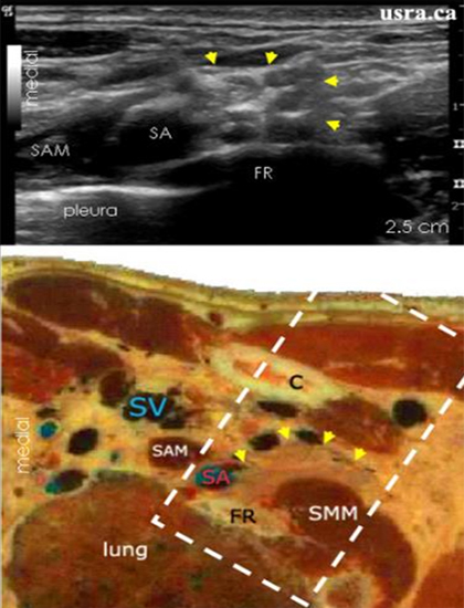
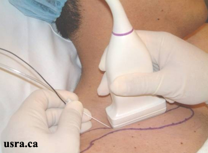
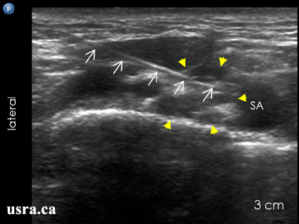
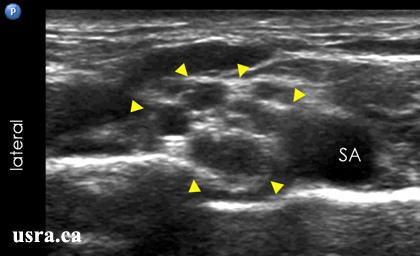
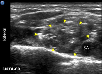
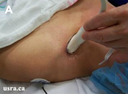
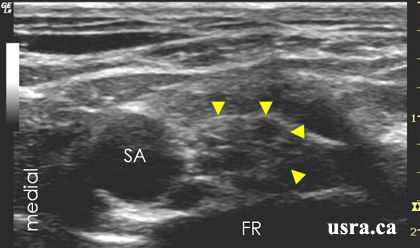
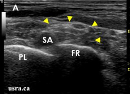
Figure A shows transducer in proper position. The structures of interest are clearly visualized.

Arrowheads = brachial plexus FR = first rib PL = pleura SA = subclavian artery

Figure B shows that the transducer is angled too anteriorly. None of the structures of interest are clearly visualized.

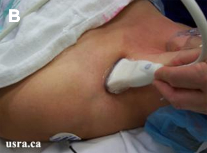
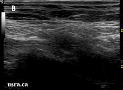
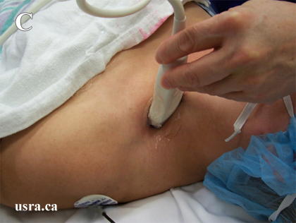
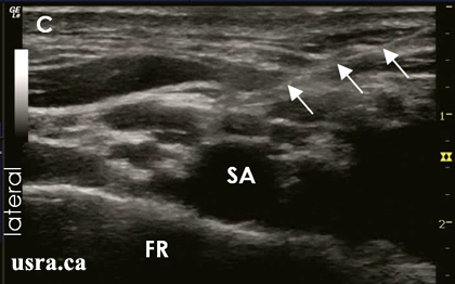
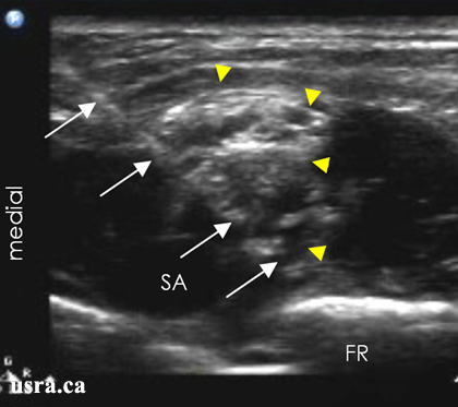
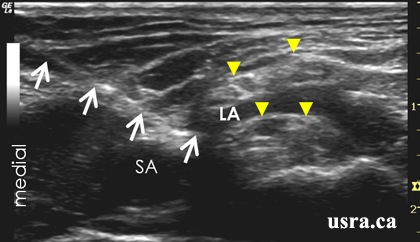
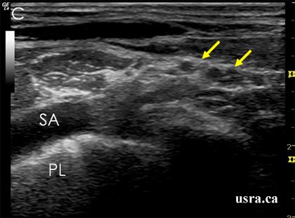
Figure C shows that the transducer is angled too posteriorly. The subclavian artery and the brachial plexus are now visualized oblique and the view is not optimal.

Arrowheads = brachial plexus SA = subclavian artery PL = pleura

2. Suprascapular Artery and Transverse Cervical Artery

Hypoechoic vessels may be seen in transverse or longitudinal section among the nerve trunks/divisions in the supraclavicular region. Most common is the suprascapular artery or the transverse cervical artery. Because of similarity in appearance (both hypoechoic), it is important to differentiate the vascular structures from the nerve structures by using color Doppler. This is crucial to avoid targeting small arteries mistaken as nerves.

Hypoechoic nodular structures in the supraclavicular region that resemble nerves (arrowheads).

Arrowheads = nerves
SA = subclavian artery
Color Doppler demonstrates that one of the hypoechoic nodules is a small artery (blue).

SA = subclavian artery

Needle Insertion

1. In Plane Medial to Lateral Approach

This is a useful alternative and some believe that this is a safer approach because the needle is pointing away from the subclavian artery and the thorax.

However, the subclavian artery can often obstruct access to the medial and inferior portion of the nerve trunks or divisions of the brachial plexus. It may be necessary to use the needle to move the artery out of its path of penetration in some cases.

Pre Injection
Arrowheads = nerves
SA = subclavian artery
FR = first rib

Post Injection
Medial to Lateral Needle Approach
Arrows = block needle
Arrowheads = nerves
SA = subclavian artery
LA = local anesthetic

2. Oblique Posterior to Anterior Approach

The transducer held in an oblique plane can image the brachial plexus in 2 dimensions:

1) medial to lateral
2) anterior to posterior

As opposed a single medial to lateral orientation when the transducer is held parallel to the clavicle. The oblique angle can enhance nerve visualization because the brachial plexus is located both lateral and posterior to the subclavian artery.

Catheter Insertion

  • Continuous supraclavicular block (CSCB) is indicated for arm and hand analgesia (see Catheter Technique).
  • The in plane needle insertion approach is recommended for CSCB. It is of paramount importance to visualize the needle tip at all times to ensure placement above the first rib and pleura.
  • The needle may be inserted in a medial to lateral direction or vice versa. The medial to lateral insertion direction has the theoretical advantage of added safety since the needle is pointing away from the thorax and towards the shoulder.
  • The block needle is advanced to the space immediately above the first rib and lateral to the subclavian artery.
  • Injection of local anesthetic or D5W solution (if nerve stimulation is desired) through the needle to distend the supraclavicular region is recommended to facilitate the ease of catheter advancement.
  • Local anesthetic spread can be observed in real time during catheter injection.
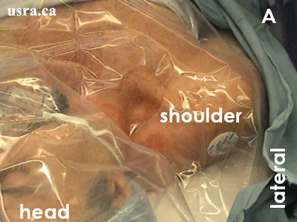
A. Patient preparation and sterile draping 
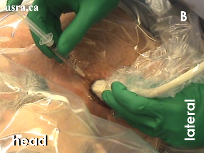
B. Pre-block scanning and local anesthetic skin infiltration
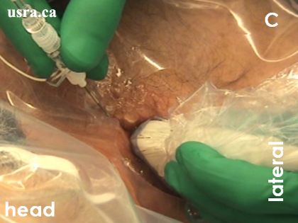
C. Block needle advancement (medial to lateral) under ultrasound guidance 
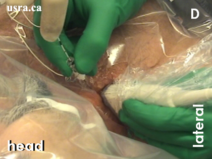
D. Catheter insertion with the help of an assistant
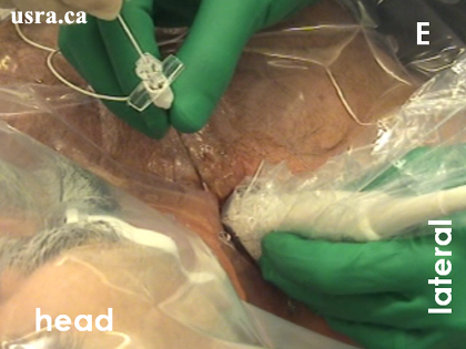
E. Removal of block needle 
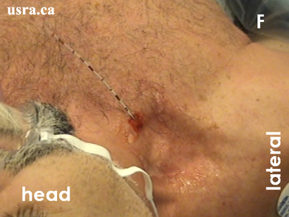
F. Catheter exits above the clavicle
A. Pre-block Scan

Arrowheads = brachial plexus trunks/divisions
FR = first rib
SA = subclavian artery
B. The block needle (arrows) is inserted in a medial to lateral direction to reach the lateral corner of the subclavian artery (SA) and above the first rib (FR).
C. The catheter (arrows) is now visualized in long axis after removal of the block needle. It is difficult to accurately identify the catheter tip.

FR = first rib
SA = subclavian artery
D. Although the catheter tip may not be seen easily, local anesthetic spread observed within the subclavian sheath compartment (arrowheads) indicates proper catheter tip location.

FR = first rib
SA = subclavian artery

Image Gallery

Pre Injection Scan
This is a suboptimal view of the brachial plexus. The nerve trunk above the first rib is not clearly seen.

SA = subclavian artery
FR = first rib
Pre Injection Scan
The transducer position is now adjusted compared to previous to visualize the nerves lying lateral to the subclavian artery above the first rib (FR). All the nerve trunks, first rib and subclavian artery (SA) are clearly visualized.
Pre Injection Scan
The needle (arrows) is inserted in plane with the ultrasound beam. Note that the needle tip is above the first rib (FR). As the needle is slowly advanced towards the first rib, inject a small volume of local anesthetic (1 mL) to hydro dissect the fascial sheath and perineural tissues. Fluid injection will indicate the needle tip location.

SA = subclavian artery
Post Injection Scan
The entire nerve compartment (fascial sheath, arrowheads) is now enlarged after injection. Scan the nerves cephalad and caudad to assess the extent of local anesthetic spread.

SA = subclavian artery
FR = first rib

2. Local Anesthetic Spread Pattern

A. Local anesthetic (LA) is visualized outside the plexus sheath.

Arrowheads = nerve trunks
FR = first rib
PL = pleura
SA = subclavian artery
B. Local anesthetic (LA) is visualized within the expanded plexus sheath.

Arrowheads = nerve trunks
FR = first rib
PL = pleura
SA = subclavian artery

Video Gallery

Selected References

  • Chan VW, Perlas A, Rawson R, Odukoya O: Ultrasound-guided supraclavicular brachial plexus block. Anesth Analg 2003; 97: 1514-7
  • Perlas A, Chan VW, Simons M: Brachial plexus examination and localization using ultrasound and electrical stimulation: a volunteer study. Anesthesiology 2003; 99: 429-35
  • Demondion X, Herbinet P, Boutry N, Fontaine C, Francke JP, Cotten A: Sonographic mapping of the normal brachial plexus. Am J Neuroradiol 2003; 24: 1303-9
  • Williams SR, Chouinard P, Arcand G, Harris P, Ruel M, Boudreault D, Girard F: Ultrasound guidance speeds execution and improves the quality of supraclavicular block. Anesth Analg 2003; 97: 1518-23
  • Martinoli C, Bianchi S, Santacroce E, Pugliese F, Graif M, Derchi LE: Brachial plexus sonography: a technique for assessing the root level. Am J Roentgenol 2002; 179: 699-702
  • Apan A, Baydar S, Yilmaz S, Uz A, Tekdemir I, Guney S, Elhan A: Surface landmarks of brachial plexus: ultrasound and magnetic resonance imaging for supraclavicular approach with anatomical correlation. Eur J Ultrasound 2001; 13: 191-6
  • Sheppard DG, Iyer RB, Fenstermacher MJ: Brachial plexus: demonstration at US. Radiology 1998; 208: 402-6
  • Yang WT, Chui PT, Metreweli C: Anatomy of the normal brachial plexus revealed by sonography and the role of sonographic guidance in anesthesia of the brachial plexus. Am J Roentgenol 1998; 171: 1631-6
  • Kapral S, Krafft P, Eibenberger K, Fitzgerald R, Gosch M, Weinstabl C: Ultrasound-guided supraclavicular approach for regional anesthesia of the brachial plexus. Anesth Analg 1994; 78: 507-13

Share to Facebook Share to Twitter More...