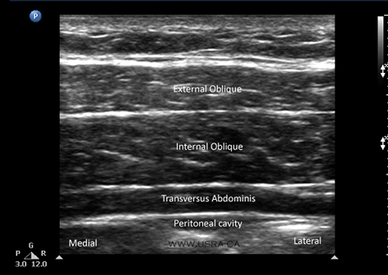
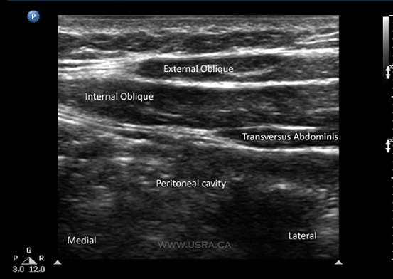
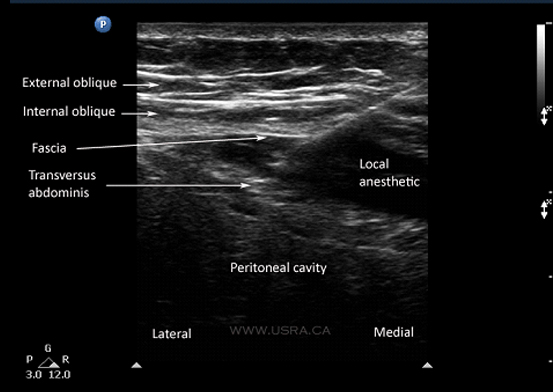
Youtube

Advancing the Science of Ultrasound Guided Regional Anesthesia and Pain Medicine

Transversus Abdominis Plane (TAP) Block

General

The transversus abdominis plane (TAP) block was first described as a landmark-guided technique involving needle insertion at the triangle of Petit.1,2 This is an area bounded by the latissimus dorsi muscle posteriorly, the external oblique muscle anteriorly and the iliac crest inferiorly (the base of the triangle). A needle is inserted perpendicular to all planes, looking for a tactile endpoint of two pops. The first pop indicates penetration of the external oblique fascia and entry into the plane between external and internal oblique muscles; the second pop signifies entry into the TAP plane between internal oblique and transversus abdominis muscles. It has been shown to provide good postoperative analgesia for a variety of procedures.3-5

More recently, ultrasound-guided techniques of TAP block have been described.6-8 A variation of the classic TAP block, the subcostal TAP block, has also been described; it is designed to provide more reliable coverage of the upper abdominal wall.9 More information on this block can be found at www.heartweb.com.au.

Anatomy

The anterior abdominal wall (skin, muscles, parietal peritoneum) is innervated by the anterior rami of the lower 6 thoracic nerves (T7 to T12) and the first lumbar nerve (L1). Terminal branches of these somatic nerves course through the lateral abdominal wall within a plane between the internal oblique and transversus abdominis muscles (Figure 1). This intermuscular plane is called the transversus abdominis plane (TAP). Injection of local anesthetic within the TAP can therefore potentially provide unilateral analgesia to the skin, muscles, and parietal peritoneum of the anterior abdominal wall from T7 to L1.2

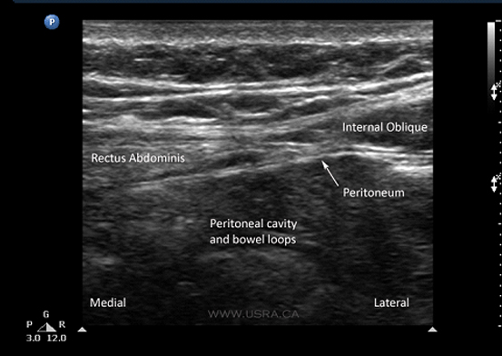
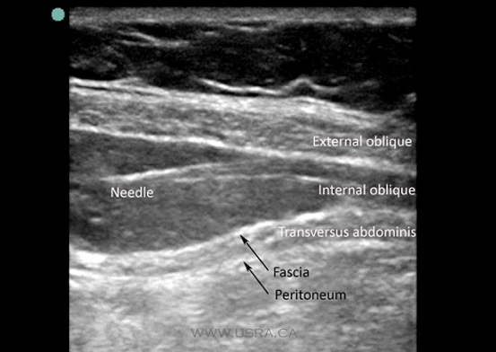
A recent cadaveric study of the TAP anatomy revealed the following points, which are pertinent to performance of the TAP block.10

  1. There is a fascial sheath between the internal oblique and transversus abdominis muscles. The nerves lie deep to this fascia.
  2. Nerves of T6-T9 enter the TAP medial to the anterior axillary line. T6 enters the TAP just lateral to the linea alba, and T7-T9 at progressively increasing distances from the linea alba. Nerves running in the TAP lateral to the anterior axillary line,on the other hand, originate from segmental nerves T9-L1. This may explain the observation of some authors that the TAP block is only suitable for lower abdominal surgery.9,11
  3. There is extensive branching and communication of the segmental nerves in the TAP. In particular the T9-L1 branches form a so-called "TAP plexus" that runs with the deep circumflex iliac artery. This may partly account for the ability of a single injection to cover several segmental levels.

Figure 1 Anatomy of the transversus abdominis plane (TAP).

Scanning Technique

  • The ultrasound guided TAP block is considered a BASIC skill level block. It is relatively simple to identify the fascial plane between the internal oblique and transversus abdominis muscles.
  • The patient is placed in a supine position and the abdomen is exposed between the costal margin and the iliac crest.
  • A linear, high-frequency transducer is recommended for this block, as the relevant anatomical structures are relatively shallow.
  • Following skin and transducer preparation, the transducer is placed in an axial (transverse) plane, above the iliac crest, and in the region of the anterior axillary line.
  • The terminal branches of the anterior rami of T7 to L1 cannot be visualized but are expected to lie within the TAP between internal oblique and the transverse abdominis muscles above the iliac crest.
  • Identify the three muscular layers of the abdominal wall: the external oblique (most superficial), the internal oblique and transversus abdominis muscles (Figure 2). Among the three muscles, the internal oblique muscle is usually the most prominent layer. In the lower medial aspect of the abdominal wall, the external oblique muscle gives way to the external oblique aponeurosis and may therefore appear as a layer of fascia instead of muscle.
  • The peritoneal cavity lies deep to the transversus abdominis muscle layer and may be identified by the peristaltic movements of bowel loops.
Figure 2 Muscular layers of the anterolateral abdominal wall.
 

If there is difficulty in distinguishing the three muscle layers, it is helpful to start the scan in the midline over the rectus abdominis muscle. The rectus abdominis muscle is the only muscular layer in the midline (Figure 3).

Figure 3 Muscular layers of the anteromedial abdominal wall, near the midline.
 

The rectus abdominis muscle tapers laterally to a junction that leads to the three muscle layers of the lateral abdominal wall (Figures 3 and 4). The internal oblique, transversus abdominis and intervening TAP are easily identified at this point, and can be traced laterally to the region above the iliac crest where the block is to be performed.

Figure 4 Muscular layers of the anterior abdominal wall beyond the lateral border of rectus abdominis.
 

It is common to visualize small vessels within the TAP. The color Doppler function may be used to confirm vascular identity.

Anatomical Correlation

Coming Soon

Nerve Localization

It is not possible to visualize branches of the thoracolumbar nerves in the TAP plane.

Needle Insertion Approach

  • The ultrasound guided TAP block is considered a BASIC skill level block. It is relatively simple to identify the fascial plane between the internal oblique and transversus abdominis muscles.

In Plane Approach

  • An 80-120 mm 22 G short beveled block needle is inserted in-plane with the transducer, in an anterior-posterior direction (Figure 5).
  • Choosing an insertion point some distance away from the transducer permits a shallower needle trajectory and thus improves needle shaft and tip visualization.
  • Alternatively, a spinal needle or Tuohy needle may be used and connected to the syringe via short extension tubing.
Figure 5 In-plane TAP block.
 

In patients with a protuberant abdomen, manual retraction of the abdominal wall by an assistant is a useful maneuver to facilitate needle insertion (Figure 6).

Figure 6 Manual retraction of abdominal wall by assistant to facilitate in-plane TAP block.
 

It is important to deposit local anesthetic deep to the fascial layer that separates the internal oblique and transversus abdominis muscles (see Anatomy). Accurate placement of the needle tip may be facilitated by injection of a small amount of fluid (1-2 mL of saline or local anesthetic) to "hydro dissect" the appropriate plane.


Out of Plane Approach

Coming Soon

Local Anesthetic Injection

  • Correct needle tip position and deposition of local anesthetic is indicated by the appearance of a hypoechoic fluid pocket immediately deep to the hyperechoic fascial plane below the internal oblique, and above the transversus abdominis (Figure 7).
Figure 7 In-plane injection of local anesthetic into the TAP plane, between the transversus abdominis muscle and the fascial layer deep to the internal oblique muscle.
 
  • If the needle tip is intramuscular instead of in the correct plane, a pattern of fluid spread consistent with intramuscular fluid injection will be seen instead.
  • A total of 20-30 mL of local anesthetic (e.g., ropivacaine 0.5 to 0.75%) is injected into this plane on each side. The maximum recommended dose of local anesthetic (3 mg/kg of ropivacaine) should not be exceeded.
  • During local anesthetic injection, it is advisable to scan the abdomen cephalad and caudad to determine the extent of longitudinal spread. Medial and lateral scanning will determine the extent of horizontal spread.

Clinical Pearls

Coming Soon

Catheter Insertion

Coming Soon

Image Gallery

Figure 8 Injection above the fascial plane separating the internal oblique and transversus abdominis muscles. (Image courtesy of Dr. Victor Chee)

Video Gallery

Selected References

  • Rafi AN. Abdominal field block: a new approach via the lumbar triangle. Anaesthesia 2001;56(10):1024.
  • McDonnell JG, O'Donnell BD, Farrell T, et al. Transversus abdominis plane block: a cadaveric and radiological evaluation. Reg Anesth Pain Med 2007;32(5):399.
  • Carney J, McDonnell JG, Ochana A, et al. The transversus abdominis plane block provides effective postoperative analgesia in patients undergoing total abdominal hysterectomy. Anesth Analg 2008;107(6):2056.
  • McDonnell JG, Curley G, Carney J, et al. The analgesic efficacy of transversus abdominis plane block after cesarean delivery: a randomized controlled trial. Anesth Analg 2008;106(1):186.
  • McDonnell JG, O'Donnell B, Curley G, et al. The analgesic efficacy of transversus abdominis plane block after abdominal surgery: a prospective randomized controlled trial. Anesth Analg 2007;104(1):193.
  • Hebbard P, Fujiwara Y, Shibata Y, et al. Ultrasound-guided transversus abdominis plane (TAP) block. Anaesth Intensive Care 2007;35(4):616.
  • Tran TM, Ivanusic JJ, Hebbard P, et al. Determination of spread of injectate after ultrasound-guided transversus abdominis plane block: a cadaveric study. Br J Anaesth 2009;102(1):123.
  • Walter EJ, Smith P, Albertyn R, et al. Ultrasound imaging for transversus abdominis blocks. Anaesthesia 2008;63(2):211.
  • Hebbard P. Subcostal transversus abdominis plane block under ultrasound guidance. Anesth Analg 2008;106(2):674.
  • Rozen WM, Tran TM, Ashton MW, et al. Refining the course of the thoracolumbar nerves: a new understanding of the innervation of the anterior abdominal wall. Clin Anat 2008;21(4):325.
  • Shibata Y, Sato Y, Fujiwara Y, et al. Transversus abdominis plane block. Anesth Analg 2007;105(3):883; author reply 883.

Share to Facebook Share to Twitter More...