Youtube

Advancing the Science of Ultrasound Guided Regional Anesthesia and Pain Medicine

Sciatic Nerve Block - Gluteal Region

Anatomy

The sciatic nerve is the largest nerve in the human body, originating from the lumbosacral plexus (L4-5 and S1-3) and providing sensory and motor innervation to the lower extremity. The sciatic nerve exits the pelvis via the greater sciatic foramen below the piriformis muscle. In the gluteal region, the sciatic nerve courses between muscle layers. The nerve is deep (anterior) to the gluteus maximus muscle and is superficial (posterior) to the inner muscle layers (superior and inferior gemellus muscles, obturator internus muscle, quadratus femoris muscle).

It courses down the midline of the posterior thigh and branches into the tibial and common peroneal nerves usually in the popliteal fossa. Sciatic nerve block is most commonly performed for foot or ankle surgery.

Posterior view of the gluteal region showing the relationship of the sciatic nerve to surrounding muscle layers:

1 = sciatic nerve
2 = piriformis muscle
3 = gluteus maximus muscle
4 = inner muscle layer (superior and inferior gemellus muscles, obturator internus muscle, quadratus femoris muscle)

Transverse View of the Sciatic Nerve at the Ischial Spine Region


Posterior View of The Gluteal Region


The relationship of the sciatic nerve to surrounding bony structures. The red dotted box marks the target of the sciatic nerve for the Labat approach at the ischial spine level.

At the level of the ischial spine, the sciatic nerve lies on top of the ischial bone and lateral and posterior to the ischial spine. Important vascular landmarks medial to the sciatic nerve and immediately next to the ischial spine are the pudendal vessels (artery and vein).

Scanning Technique

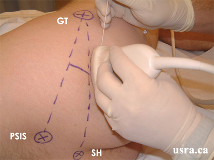
  • Position the patient semi-prone (Sims' position) with the hip and knee flexed and the operative side uppermost.
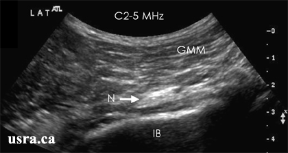
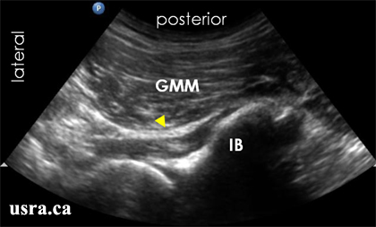
  • After skin and transducer preparation (see transducer preparation section), place a curved low frequency 2-5 MHz transducer firmly on the buttock region to capture the best possible transverse view of the sciatic nerve.
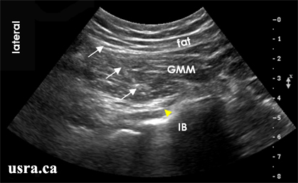
  • Optimize machine imaging capability. Select appropriate depth of field (usually > 4 cm from the skin surface), focus range and gain.
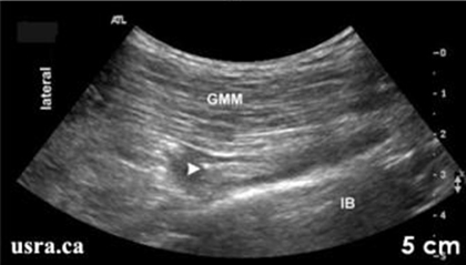
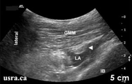
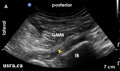
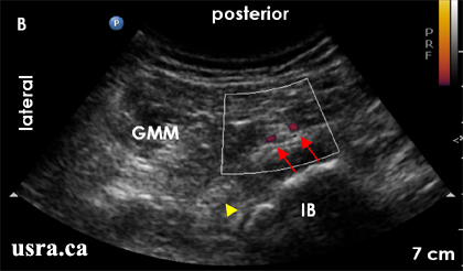
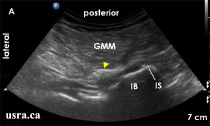
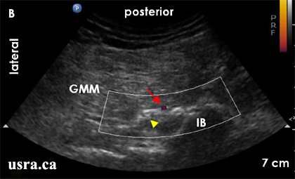
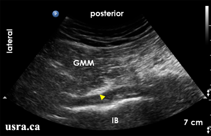
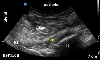
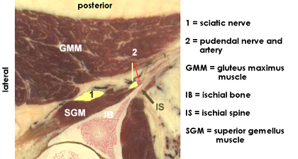
  • Observe on the screen the transverse view of the sciatic nerve (short axis). The sciatic nerve in the gluteal region appears predominantly hyperechoic (bright) and is often wide and flat in the transverse view on ultrasound.
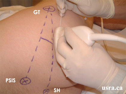
Transducer over right gluteal region.

GT = greater trochanter
PSIS = posterior inferior iliac spine
SH = sacral hiatus

Anatomical Correlation

Lateral

Arrow = sciatic nerve
GMM = gluteus maximus muscle
IB = ischial bone
PN and PV = pudendal nerve and vessels
SN = sciatic nerve

Nerve Localization

  • Perform a systematic anatomical survey from cephalad to caudad and from superficial to deep.
  • First identify the bony structures that are adjacent to the sciatic nerve. Locate the ischial bone (a hyperechoic line with the bony shadow underneath). Locate the widest portion of the ischial bone and the ischial spine medially.
The bony segment at the level of the ischial spine (A) is the widest compared to the bony segments cephalad (B) and caudad (C).
  • Locate the bulky gluteus maximus muscle (GMM) that is superficial and posterior to the sciatic nerve. The sciatic nerve is expected to lie between the GMM and ischial bone.
  • May use color Doppler to locate the pudendal vessels (artery and vein) that are adjacent to the ischial spine, all medial to the sciatic nerve.
  • Another vascular structure that may be noted immediately adjacent to the sciatic nerve is the inferior gluteal artery.
  • Angle the transducer slightly cephalad or caudad to capture the best possible transverse view of the sciatic nerve.
The bony segment at the level of the ischial spine (A) is the widest compared to the bony segments cephalad (B) and caudad (C).

Arrowhead = sciatic nerve
GMM = gluteus maximus muscle
IB = ischial bone
  • Locating the sciatic nerve in this region can be challenging because of the required depth of penetration especially when there is an abundant amount of overlying adipose tissue.
  • The sciatic nerve can be wide but thin. It can be difficult to separate the thin sciatic nerve from the overlying gluteus maximus muscle layer in the transverse view.
  • Follow the nerve by scanning proximally (cephalad) and distally (caudad) to follow the course of the nerve. It may be necessary to first identify the nerve in the subgluteal region and then trace the nerve proximally should visualization be difficult.

Needle Insertion Approach

  • Ultrasound guided sciatic nerve block in the gluteal region is considered an ADVANCED skill level block.
  • The sciatic nerve may be difficult to visualize in this region because of the required depth of beam penetration and lower image resolution from the use of a lower frequency transducer. The overlying adipose tissue in the buttock may be sizable, the sciatic nerve may be quite flat in the transverse view and the required angle of needle penetration may be quite steep.

In Plane Approach

For the In Plane approach, insert a 8 cm 22G insulated block needle on the outer (lateral) end of the ultrasound transducer after skin local anesthetic infiltration. Advance the needle along the long axis of the transducer in the same plane as the ultrasound beam. In this way, the needle can be visualized in real time at the time of needle advancement towards the target nerve.
  • Advance the needle through the gluteus maximus muscle to reach the sciatic nerve above the ischial bone.
  • Because of the depth and the angle of needle penetration (often steep, > 45 degrees), it is often not possible to accurately visualize the needle shaft and tip. Most commonly, needle and tissue movements are observed during needle advancement.
  • Nerve movement may be observed upon needle contact. Additionally electrical stimulation is recommended to confirm needle to nerve contact.
  • Observe the pattern of local anesthetic spread around the target nerve in real time during injection. One may adjust needle position half way during injection to optimize local anesthetic spread.

In Plane Needle Approach to the Gluteal Sciatic Nerve

Arrows = block needle
Arrowhead = sciatic nerve
GMM = gluteus maximus muscle
IB = ischial bone

Out of Plane Approach

  • For the OOP approach, align the nerve target at the midpoint of the transducer and then insert the block needle in the same location.
  • The OOP approach is often used for catheter insertion.
  • For both IP and OOP approaches, scanning prior to needling will determine the angle, distance and depth of needle penetration.
  • Observe tissue and needle movement as the needle is advanced towards the target penetration.
  • Clear identification of the needle tip can be technically challenging.
  • Confirm needle to nerve contact by electrical stimulation and observe local anesthetic spread.

Local Anesthetic Injection

  • Aim to deposit local anesthetic around the sciatic nerve circumferentially.
  • However, this may be challenging because the sciatic nerve in this region may be flat and thin in cross section. It is common to find local anesthetic on only one side of the nerve after a single injection.
  • Injection to the other side of the nerve may require a second needle insertion site. A hypoechoic (fluid) expansion can be seen during local anesthetic injection.
  • Expansion in the gluteus maximus muscle indicates superficial intramuscular injection. Advance the needle accordingly.
  • Inject 15-20 mL of local anesthetic for postoperative analgesia.
Pre Injection

Arrowhead = sciatic nerve
GMM = gluteus maximus muscle
IB = ischial bone
Post Injection

Arrowhead = sciatic nerve
GMM = gluteus maximus muscle
IB = ischial bone
LA = local anesthetic (deep to the nerve)

Clinical Pearls

Nerve Localization

1. Identification of the Pudendal Vessels


The pudendal vessels are vascular landmarks at the level of the ischial spine. The vessels are usually visualized adjacent to the ischial spine and medial to the sciatic nerve. It is possible to use Color Doppler or Color Power Doppler to identify the pudendal vessels (artery and vein).

A. Baseline Scan

Arrowhead = sciatic nerve
GMM = gluteus maximus muscle
IB = ischial bone
B. Scan with Color Power Doppler

Arrowhead = sciatic nerve
GMM = gluteus maximus muscle
IB = ischial bone
Red arrow = pudendal artery and vein

2. Identification of the Inferior Gluteal Artery

Another vascular landmark at the level of the ischial spine is the inferior gluteal artery. This structure is usually visualized adjacent to the sciatic nerve. This small artery is more lateral than the pudendal artery.

A. Baseline Scan

Arrowhead = sciatic nerve
GMM = gluteus maximus muscle
IB = ischial bone
IS = ischial spine
B. Scan with Color Power Doppler

Arrowhead = sciatic nerve
GMM = gluteus maximus muscle
IB = ischial bone
Red arrow = inferior gluteal artery

Catheter Insertion

Catheter is not commonly inserted at this proximal location for continuous sciatic nerve block.

Image Gallery

Pre-injection Scan at the Level of the Ischial Spine

Arrowhead = sciatic nerve
GMM = gluteus maximus muscle
IB = ischial bone

Pre-injection Scan BELOW the Level of the Ischial Spine

Arrowhead = sciatic nerve
GMM = gluteus maximus muscle
HJ = hip joint
IB = ischial bone

Post-injection Scan at the Level of the Ischial Spine

Arrowhead = sciatic nerve
GMM = gluteus maximus muscle
IB = ischial bone
LA = local anesthetic

Video Gallery

Selected References

  • Moayeri N, van Geffen GJ, Bruhn J, Chan VW, Groen GJ. Correlation among ultrasound, cross-sectional anatomy, and histology of the sciatic nerve: a review. Reg Anesth Pain Med 2010;35:442-9.
  • Saranteas T, Chantzi C, Paraskeuopoulos T, Alevizou A, Zogojiannis J, Dimitriou V, Kostopanagiotou G: Imaging in Anesthesia: The Role of 4 MHz to 7 MHz Sector Array Ultrasound Probe in the Identification of the Sciatic Nerve at Different Anatomic Locations. Reg Anesth Pain Med 2007; 32: 537-8
  • Chantzi C, Alevizou A, Saranteas T, Zogogiannis J, Iatrou C, Dimitriou V: Usefulness of the two to 5 MHz ultrasound probe in examination and block of the sciatic nerve in orthopedic trauma patients: a preliminary study. J Clin Anesth 2007; 19: 486-8
  • Chan VW, Nova H, Abbas S, McCartney CJ, Perlas A, Quan XD: Ultrasound Examination and Localization of the Sciatic Nerve: A Volunteer Study. Anesthesiology 2006; 104: 309-14
  • Graif M, Seton A, Nerubai J, Horoszowski H, Itzchak Y: Sciatic nerve: sonographic evaluation and anatomic-pathologic considerations. Radiology 1991; 181: 405-8
  • Hullander M, Spillane W, Leivers D, Balsara Z: The use of Doppler ultrasound to assist with sciatic nerve blocks. Reg Anesth. 1991; 16: 282-4

Share to Facebook Share to Twitter More...