Youtube

Advancing the Science of Ultrasound Guided Regional Anesthesia and Pain Medicine

Inguinal Canal Block

Introduction

Blockade of the genital branch of the genitofemoral nerve (gGbFN) in the inguinal canal (ICAN) may be performed in conjunction with ilioinguinal and iliohypogastric nerve block to provide surgical anesthesia for inguinal herniorrhaphy. Inguinal canal block is also called "spermatic cord block" in male. It is performed as a diagnostic block to define the etiology of pain in the scrotum, testicle, labia majora and vagina that may be attributed to genitofemoral neuropathy.

Anatomy

The genitofemoral nerve (GFN), originating from L1 and L2 part of the lumbar plexus, is the smallest nerve of the plexus. After emerging from its origin, the GFN pierces the psoas major muscle at its ventral surface. The nerve may emerge as a single nerve or more often it is already divided into 1) the genital branch (gbGFN) and 2) the femoral branch (fbGFN). The gbGFN enters and courses within the inguinal canal (ICAN).

In males, the gbGFN is situated between the cremaster muscle and internal spermatic fasciae. The genital branch provides innervation to the cremaster muscle and the scrotal skin. Terminal sensory branches of the gbGFN may innervate part of the upper, inner, and medial thigh.

In females, the gbGFN provides sensation to the mons pubis and labia majora and the terminal sensory branches innervates part of the upper, inner, and medial thigh. The femoral branch provides innervation to the skin over the femoral triangle.

The inguinal canal is a cylindrical passage in the lower part of the anterior abdominal wall that runs obliquely anterior, inferior and medial (Figure 1). The inguinal canal is bounded by the aponeurosis of the external oblique muscle anteriorly (white arrows), the transversalis fascia posteriorly (purple arrows), the lower border of the transversus abdominis muscle cranially (brown arrows) and the inguinal ligament caudally (green arrow). Immediately outside and posterior to the inguinal canal is the inferior epigastric artery (IEA, red asterisk), a branch of the external iliac artery (EIA).

Figure 1. A parasagittal cross section of the lower abdominal wall showing the inguinal canal surrounded by arrows

* = offspring of the inferior epigastric artery
EIA = external iliac artery
EIV = external iliac vein
white arrow = aponeurosis of the external oblique muscle anteriorly
purple arrow = transversalis fascia posteriorly
brown arrow = lower border of the transversus abdominis muscle cranially
green arrow = inguinal ligament caudally

Movie 1. A parasagittal cross section of the inguinal canal


Inside the inguinal canal are 2 nerves, the genital branch of the genitofemoral nerve (gGbFN) and the ilioinguinal nerve (Figure 2). It also contains the spermatic cord in men with its three fascial layers and the round ligament of the uterus in women. The spermatic cord is enveloped by 3 fascial layers- the external spermatic fascia, the cremasteric fascia (CF) and the internal spermatic fascia (ISF). The gbGFN is inside the spermatic cord (usually in a duplication of the CF), while the ilioinguinal nerve runs inside the ICAN but outside the spermatic cord. Note that the ilioinguinal nerve is significantly larger in size than the gbGFN. Within the ISF are the vas deferens (with its small accompanying artery), the testicular artery, and the pampiniform venous plexus.

Figure 2A. Anatomy of the inguinal canal


The red box indicates the inguinal canal to be magnified.

Figure 2B. Magnification of the inguinal canal as outlined by the red box

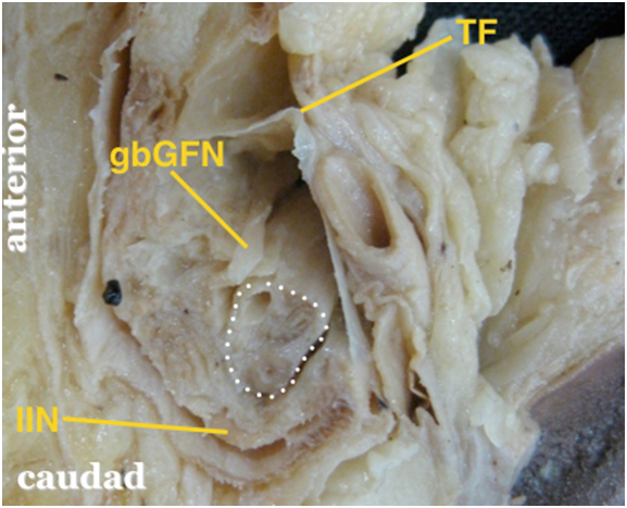
The white dotted area outlines the internal spermatic fascia of the spermatic cord containing the vas deferens, testicular artery and pampiniform plexus.
gbGFN = genital branch of the genitofemoral nerve within the cremasteric facia (external spermatic fascia removed)
IIN = ilioinguinal nerve which runs within the inguinal canal but outside the spermatic cord; note that the IIN is significantly bigger than the gbGFN
TF = transversalis fascia

Movie 2. Magnification of the inguinal canal

Sonoanatomy

Sonographically, the inguinal canal is an oval shaped structure with a predominantly hyperechoic appearance, as a result of the fascial layers of the spermatic cord and its contents. The small hypoechoic "dots" within the inguinal canal represent mainly the testicular artery, pampiniform venous plexus, vas deferens. In the longitudinal view, the inguinal canal containing the spermatic cord is outlined by its fasciae and surrounding structures (Figure 3).

Figure 3. Longitudinal view of the inguinal canal

APO = anterior aponeurosis of external oblique muscle
EIA = external iliac artery
FA = femoral artery
IC = inguinal canal
IEA = inferior epigastric artery posterior to the inguinal canal
TF = transversalis fascia

Scanning Technique

Position the patient spine.

After skin and transducer preparation, place a linear high frequency 10-12 MHz transducer longitudinally on the skin surface to obtain a long axis view of the inguinal canal (Figure 4).

Optimize machine imaging capability by selecting the appropriate depth of field (usually within 1-2 cm), focus range and gain.

The spermatic cord within the inguinal canal appear predominantly hyperechoic with some internal hypoechoic "dots".

Figure 4. Longitudinal transducer position in the inguinal region to capture a long axis view of the inguinal canal


Movie 3. Transducer position for scanning the inguinal canal

Canal Localization

External palpable landmarks are the anterior superior iliac spine, the pubic tubercle and pulsation of the femoral artery.

Place the transducer longitudinally in the groin to capture a long axis view of the femoral artery.

Then move the transducer progressively cephalad to visualize the external iliac artery, a continuation of the femoral artery, that is located deeper (Figure 5).

Figure 5. A longitudinal scan showing the femoral artery which continues to become the external iliac artery cranially

EIA = external iliac artery
FA = femoral artery

Move the transducer medially from this point to identify the inferior epigastric artery, a branch of the external iliac artery. The inferior epigastric artery is an important landmark for locating the inguinal canal and the spermatic cord in man or the round ligament in woman (Figure 6).

Figure 6. Sonogram showing the origin of the inferior epigastric artery from the external iliac artery

EIA = external iliac artery
IEA = inferior epigastric artery

Movie 4. Localization of the inguinal canal


Moving the transducer slightly more medial will uncover the spermatic cord which is predominantly hyperechoic (Figure 7).

When the transducer is moved further medial, the external iliac artery is no longer in view and the borders of the inguinal canal are not clearly visible. However, the hyperechoic spermatic cord remains clearly visible. Figure 7. Sonogram showing the inguinal canal in cross section

EIA = external iliac artery
IEA = inferior epigastric artery
FA = femoral artery
IC = inguinal canal

Needle Insertion Approach

The inguinal canal block is considered an intermediate skill level block. Blockade within the inguinal canal can only anesthetize the genital branch of the genitofemoral nerve because the femoral branch has taken off more proximally to enter the vascular space. The major potential risk is accidental intravascular injection into the venous plexus inside the canal.

We recommend an out of plane needle approach for inguinal canal block.

After local anesthetic skin infiltration, a 22 G 5 cm needle can be inserted on either the medial or lateral side of the transducer until the needle tip reaches the inguinal canal.

One may feel a pop going through fascia.

Incremental hydrodissection is recommended along the path of needle advancement (Figure 8)

Figure 8. Sonogram showing a small fluid collection after hydro-dissection with the block needle tip anterior to the spermatic cord before local anesthetic injection


Movie 5. Injection endpoint

Local Anesthetic Injection

The goal is to inject local anesthetic inside the inguinal canal.

After advancing the needle inside the inguinal canal, 4 mL of local anesthetic is injected and then another 4 mL inside the spermatic cord. Figures 9 and 10 show the inguinal canal before and after injection respectively.

Local anesthetic injection within the inguinal canal will anesthetize the genital branch of the genitofemoral nerve and the ilioinguinal nerve. The femoral branch of the genitofemoral nerve will not be blocked because it has taken off more proximally.

Figure 9. Sonogram showing spermatic cord before injection


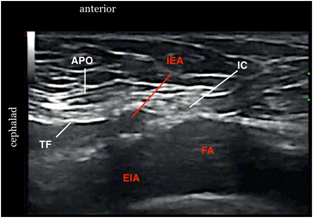
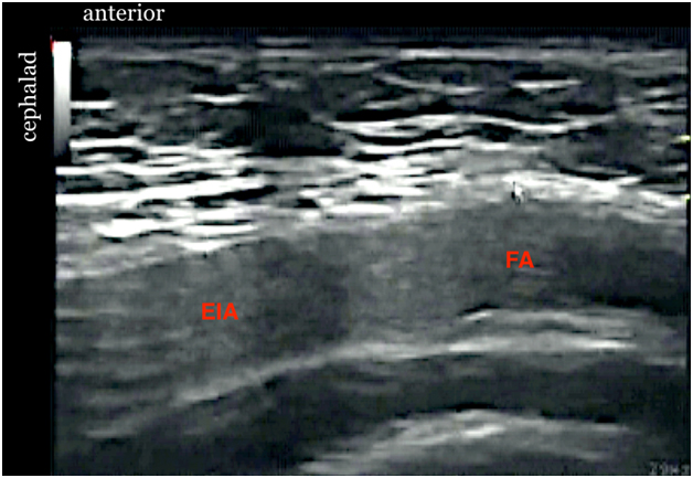
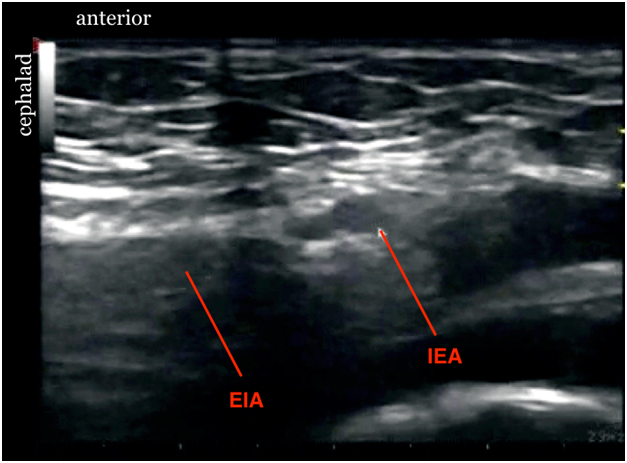
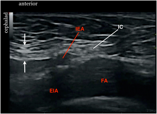
Figure 10. Sonogram showing spermatic cord after injection


Movie 6. Out of plane inguinal canal injection


Movie 7. Inguinal canal injection

Clinical Pearls

It is relatively easy to achieve an effective "inguinal canal block" without seeing the genital branch of the genitofemoral nerve (gbGFN). Local anesthetic is injected both inside and outside the spermatic cord thus blocking both the gbGFN and the ilioinguinal nerve. Thus, the gbGFN cannot be selectively blocked without affecting the ilioinguinal nerve. This is a drawback of this procedure. To selectively block the ilioinguinal nerve, one needs to approach the ilioinguinal nerve more cephalad next to the iliac crest.

There is a high degree of variability for the level at which the genitofemoral nerve divides. This will influence topographical relationships. Also, the gbGFN occupies variable locations relative to sheaths of the spermatic cord within the ICAN. The gbGFN may lie between any of the three fascial layers, within them or even outside the spermatic cord together with the ilioinguinal nerve.

A transducer of 15 MHz or higher may be required to visualize the gbGFN within the inguinal canal in slim individuals. However, because of its small size, location variability and the topographic orientation, it is highly unlikely to visualize the gbGFN clearly enough for selective blockade.

The spermatic cord may be difficult to visualize due to anisotropy or in the very obese subjects. It may be useful to change transducer angle by using the "heel in" maneuver, i.e., pressing in one end of the transducer. Another trick is to ask an assistant to grasp the spermatic cord gently between two fingers during the scan. This helps to bring the tissues closer together into a more compact structure with clearer borders.

Application of Color Doppler or Color Power Doppler will show vascular structures inside the spermatic cord.

To visualize the spermatic cord clearly, it is necessary to move the transducer medially until the external iliac artery is no longer in view. At this location, the spermatic cord may be more visible since the borders of the inguinal canal (made up of the aponeurosis of the external oblique muscle and the transversalis fasia) have mostly disappeared.

Movie 8. Transducer maneuver to overcome anisotropy

Video Gallery

Selected References

  • Bhosale PR, Patnana M, Viswanathan C, Szklaruk J. The Inguinal Canal: Anatomy and Imaging Features of Common and Uncommon Masses. Radiographics 2008;28:819-835
  • Campos NA, Chiles JH, Plunkett AR. Ultrasound-guided cryoablation of genitofemoral nerve for chronic inguinal pain. Pain Physician 2009;12:997-1000.
  • Liu WC, Chen TH, Shyu JF, et al.: Applied anatomy of the genital branch of the genitofemoral nerve in open inguinal herniorrhaphy. Eur J Surg 2002;168:145-9.
  • Peng P, Narouze S. Ultrasound-guided interventional procedures in pain medicine: A review of anatomy, sonoanatomy and procedures. Part I: Non-axial structures. Reg Anesth Pain Med 2009;34:458-474.
  • Rab M, Ebmer And J, Dellon AL: Anatomic variability of the ilioinguinal and genitofemoral nerve: Implications for the treatment of groin pain. Plast Reconstr Surg 2001;108:1618-23.
  • Shanthanna H. Successful treatment of genitofemoral neuralgia using ultrasound guided injection: a case report and short review of literature. Case Rep Anesthesiol 2014;2014:371703.
  • Tagliafico A, Bignotti B, Cadoni A, Perez MM, Martinoli C. Anatomical study of the iliohypogastric, ilioinguinal, and genitofemoral nerves using high-resolution ultrasound. Muscle Nerve 2015;51:42-8.

Share to Facebook Share to Twitter More...