Youtube

Advancing the Science of Ultrasound Guided Regional Anesthesia and Pain Medicine

Mid Humeral Block

Anatomy

The mid humeral approach was described by Dupre in 1994. The site of needle entry is at the junction between the upper 1/3 and lower 2/3 of the arm. Terminal nerves of the brachial plexus are separated at this location. The median and ulnar nerves are found superficial and adjacent to the brachial artery, the musculocutaneous nerve under the biceps muscle belly and the radial nerve posterior to the humeral shaft. The mid humeral block aims to anesthetize each nerve separately, possibly using local anesthetic with different durations of action.

Scanning Technique

  • Position the patient supine and abduct the arm to approximately 90 degrees.
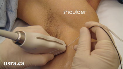
  • After skin and transducer preparation (see transducer preparation section), place a linear 38-mm, high frequency 10-12 MHz transducer firmly on the upper 1/3 of the arm to capture the best possible transverse view of the nerves in the midhumeral region.


Transducer over left midhumeral region (upper 1/3 of the arm).

  • Optimize machine imaging capability by selecting the appropriate depth of field (within 1-2 cm), focus range (usually within 1 cm) and gain.
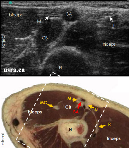
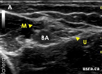
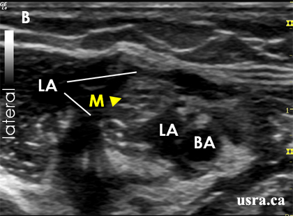
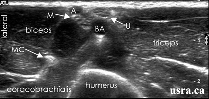
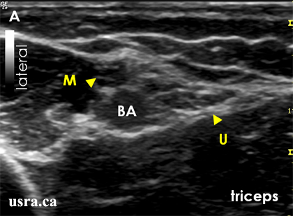
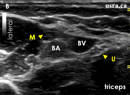
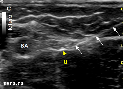
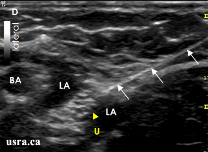
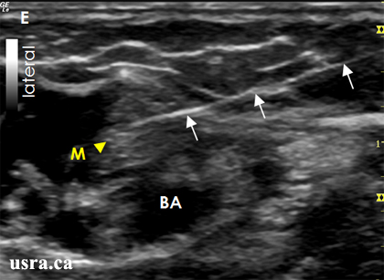
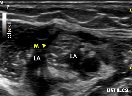
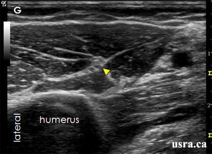
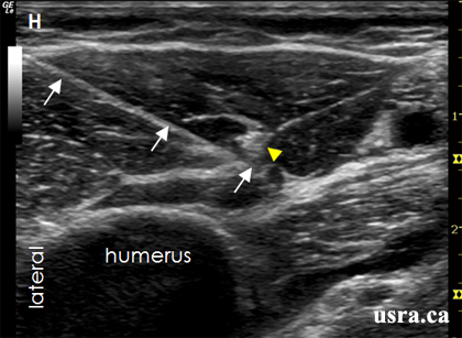
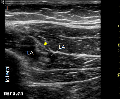
  • Visualize the median, ulnar, and musculocutaneous nerves in transverse view (short axis). Nerves in the midhumeral region have mixed echogenicity (honeycomb appearance with a mixture of hypoechoic nerve fascicles and hyperechoic connective tissues). The nerves are round or oval, and are located next to the brachial artery and vein.

Anatomical Correlation

BA = brachial artery
Box = scanned area
CB = coracobrachialis muscle
H = humerus
M = median nerve
MC = musculocutaneous nerve
R = radial nerve
U = ulnar nerve

Nerve Localization

  • Place the transducer on the upper 1/3 of the arm to obtain a transverse view of the brachial artery in an outstretched arm.
  • Relieve transducer pressure slightly on the skin to visualize the surrounding vein(s).
  • Identify the triceps, biceps and coracobrachialis muscles surrounding the artery.
  • Identify the humerus deep to the muscles.
  • The median and ulnar nerves are expected to lie superficial (often within 1 cm from the skin surface) and adjacent to the brachial artery (the median nerve lateral and the ulnar nerve medial to the artery). They often have a honey comb appearance and are heterogeneous in echogenicity.
  • Identify the musculocutaneous nerve between the biceps and coracobrachialis muscles. This nerve appears predominantly hyperechoic.
  • The radial nerve is not usually visualized at this level since it lies posterior to the humeral shaft.
  • Move the transducer proximally towards the axilla and distally towards the elbow to appreciate the course of each nerve (nerve tracing).

Needle Insertion Approach

In Plane Approach

  • Ultrasound guided midhumeral block is considered a BASIC skill level block because this is a superficial block.
  • Insert a 5 cm 22 G insulated needle parallel to the long axis of the transducer inline with the ultrasound beam (in plane approach).
  • Visualize the median, ulnar and musculocutaneous nerves in transverse view.
  • Identify the pulsatile brachial artery which is anechoic.
  • Apply firm transducer pressure to collapse surrounding venous structures.
  • The needle should be inserted at a shallow angle because the median and ulnar nerves are both superficial. As the needle travels in the same plane as the ultrasound beam, the path of advancement can be visualized in real-time as the needle approaches the target nerves.
  • Identify the nature of each individual nerve by electrical stimulation if desired.

Out of Plane Approach

Out of plane approach is an alternate approach for this block.

Local Anesthetic Injection

  • Inject 10 mL of local anesthetic at each nerve location. The minimum effective local anesthetic volume for mid humeral block has not been determined.
  • Aim to achieve circumferential spread around the median, ulnar and musculocutaneous nerves.
Figure A reveals the mid humeral region. The median nerve (M) and ulnar nerve (U) are both superficially located adjacent to the brachial artery (BA).
Figure B illustrates local anesthetic (LA) injection and circumferential spread around the median nerve (M).
The musculocutaneous nerve often requires a separate injection for blockade.

BA = brachial artery

Clinical Pearls

Nerve Localization

  • Both the ulnar and median nerves are superficial thus easy to visualize and trace proximally and distally.
  • Although the musculocutaneous nerve is deeper, it is often hyperechoic and easily found in the plane between the biceps and coracobrachialis muscles. Move the transducer laterally to bring the nerve into view.
  • The radial nerve is not visualized in this projection. It is necessary to move the transducer medially and capture the nerve in an anterior to posterior projection.

Catheter Insertion

Catheter insertion is seldom indicated for this particular block.

Image Gallery

1. Anatomical Variations

The Presence of 2 Arteries

A = a branch of BA
BA = brachial artery
M = median nerve
MC = musculocutaneous nerve
U = ulnar nerve

2. Local Anesthetic Injection Around Individual Nerves

A. Pre-injection Scan

BA = brachial artery
M = median nerve
U = ulnar nerve
B. Pre-injection Scan
The brachial vein (BV) is visualized when the transducer pressure is reduced.

BA = brachial artery
M = median nerve
U = ulnar nerve
C. A block needle (arrows) is inserted in plane to approach the ulnar nerve (U).

BA = brachial artery
D. Local anesthetic (LA) spread is observed around the ulnar nerve (U).

Arrows = the block needle
BA = brachial artery
E. The block needle (arrows) is inserted in plane to approach the median nerve (M).

BA = brachial artery
F. Circumferential local anesthetic (LA) spread is observed around the median nerve (M).

G. Pre-injection Scan

Arrowhead = musculocutaneous nerve (MC)
H. A block needle (arrows) is inserted in plane to approach the MC nerve (arrowhead).
I. Local anesthetic (LA) is observed around the MC nerve (arrowhead).

Video Gallery

Selected References

  • Perlas A, Chan V W, Simons M. Brachial plexus examination and localization using ultrasound and electrical stimulation: a volunteer study. Anesthesiology 2003; 99: 429-435.

Share to Facebook Share to Twitter More...