Youtube

Advancing the Science of Ultrasound Guided Regional Anesthesia and Pain Medicine

Gastric Ultrasound

Author

Anahi Perlas, MD, FRCPC
Associate Professor
Department of Anesthesia
University of Toronto


Biosketch

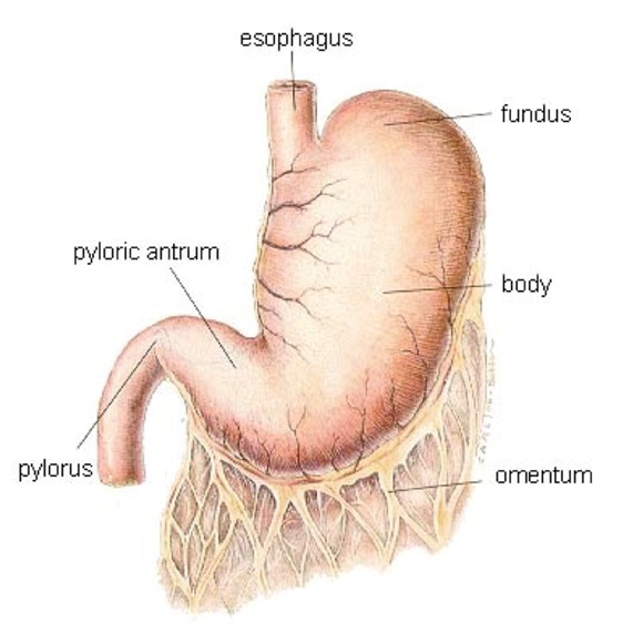
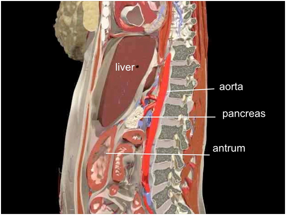
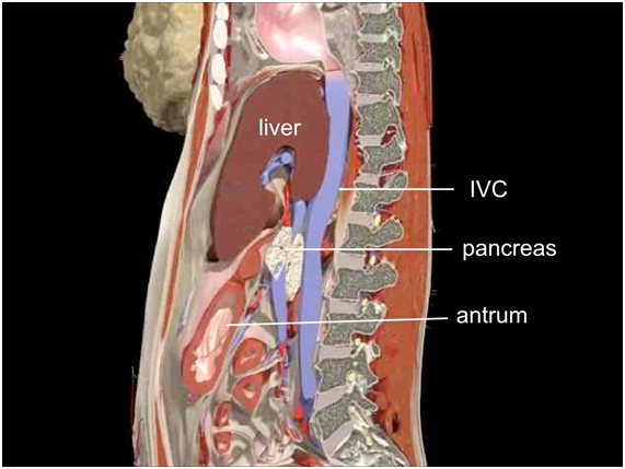
Introduction

Pulmonary aspiration of gastric content is a major anesthetic-related complication which may result in significant morbidity and mortality. This is particularly true in high risk patients and situations e.g., parturients, emergency surgery, and diseases associated with slow gastric emptying. The volume, nature (fluid vs. particulate or solid matter), and acidity of the aspirate are thought to be important factors that determine patient outcomes. Current prevention strategies rely mainly on recommended fasting periods for elective surgery i.e., NPO protocol. However, underlying medical conditions that slow gastric emptying can predispose patients to greater amount of gastric content at the time of anesthetic induction despite appropriate fasting intervals. There is a growing interest in applying bedside ultrasound as a noninvasive portable tool to assess gastric content and volume to clinically evaluate aspiration risk by providing qualitative and quantitative information.

The main objective of point-of-care (POC) gastric ultrasound is to help clinicians assess gastric contents when NPO status is unknown or uncertain in the immediate pre-anesthetic period. Other than anesthesiologists, POC gastric ultrasound is also useful to emergency physicians and intensivists who take part in sedation and airway management.

It has a number of important features:

  • it is a bedside, focused diagnostic examination
  • it is a goal-directed assessment i.e., limited in scope and target structures
  • the findings are easily recognizable
  • the scanning technique can be quickly learned and performed
  • the findings help to guide clinical care

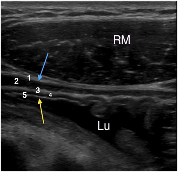
Point of care gastric ultrasound follows the I-AIM framework (I = indications, A = acquisition, I = interpretation and M = medical decision-making). This good framework describes the logical steps in performing and teaching gastric ultrasound. Acquisition of image requires selection of a proper probe (type, orientation), proper patient position, picture (image optimization) and examination protocol. Image interpretation involves interpretation of the anatomy, physiology, and artifacts. Finally, medical decision is made based on appropriate collaboration of ultrasound results with other physical examination, radiologic and laboratory test findings. It is important to acknowledge that a POC gastric ultrasound examination can conclusively rule in or rule out a clinical diagnosis. However, the ultrasound result may also be indeterminate in which case serial ultrasound evaluation is warranted.

At the present time, research is ongoing to validate the diagnostic accuracy and clinical utility of gastric ultrasound in healthy and high risk patients undergoing elective and emergency surgery.

Anatomy

There stomach has 3 anatomical parts- 1) fundus, 2) body, and 3) antrum & pylorus (Figure 1). The antrum, the most distal part of the stomach, is consistently located in the epigastric region which provides an easily accessible soft tissue window for scanning. The antrum is located inferior to the left lobe of the liver, and anterior to the pancreas, the aorta (Figure 2) and the inferior vena cava (Figure 3). It has a rather consistent shape and contains the least amount of air that blocks ultrasound beam penetration as compared to the fundus and the body of the stomach.

Figure 1. Anatomy of the Stomach

Figure 2. Anatomy of the Stomach in Relationship to the Liver and Aorta

Figure 3. Anatomy of the Stomach in Relationship to the Liver and Inferior Vena Cava

Sonoanatomy

The stomach wall is rather thick with 5 distinctive layers which can be visualized using a high-frequency linear probe with the sagittal view and the axial view (Figure 4).

From outside (external) to inside (internal), layers of the gastric wall are:

  1. serosa (thin, hyperechoic)
  2. muscularis propria (hypoechoic, usually the thickest gastric wall layer)
  3. submucosa (hyperechoic)
  4. muscularis mucosae (hypoechoic), and
  5. mucosal/lumen interface (thin hyperechoic line)

Figure 4. Axial Scan with a High Frequency Linear Transducer Showing 5 Distinct Layers of the Gastric Wall

1 = serosa (hyperechoic) (blue arrow)
2 = muscularis propria (hypoechoic)
3 = submucosa (hyperechoic)
4 = muscularis mucosa (hypoechoic)
5 = mucosal-lumen interface (hyperechoic) (yellow arrow)
Lu = stomach lumen
RM = rectus abdominis muscle

When scanned in the axial plane, both the antrum and the pylorus can be visualized as well as some posterior structures e.g., pancreas and aorta (Figure 5).

Figure 5. Axial Scan Showing Antrum and Pylorus

A = antrum
Ao = aorta
P = pancreas
Py = pylorus
RM = rectus abdominis muscle

Indications

POC gastric ultrasound is indicated when a clinician is uncertain about the patient's NPO status and /or the level of aspiration risk at the time when sedation, anesthesia or airway management is required.

Some common clinical scenarios are:

  • urgent or emergency surgical procedure without planned fasting
  • patients with co-morbidities or medical conditions that may delay gastric emptying despite adherence to appropriate NPO guidelines e.g., diabetes, neuromuscular disorders, severe renal or liver dysfunction, recent trauma, pain and opioids, active labour, and gastric dysmotility of any etiology
  • inability to obtain information on prandial status e.g., language barrier, decreased level of consciousness, and cognitive dysfunction
  • patients undergoing elective procedures with questionable or borderline adherence to fasting instructions

Image Acquisition

Gastric scanning targets the antrum which can be reliably located and identified using standard internal anatomical landmarks.

Patient Positions

  1. Scan first in the supine position as shown in Figure 6.
  2. Then scan in the right lateral decubitus position as shown in Figure 7.
  3. Occasionally, a semi-sitting position is used should the patient not able to turn to right lateral decubitus

A thorough evaluation of gastric content requires examination in the right lateral decubitus position because gastric content is expected to gravitate to the antrum in this body position. An examination performed solely in the supine position is considered incomplete and inaccurate because it underestimates the amount of gastric content. Failure to visualize gastric content in the supine position does not guarantee an empty stomach.

It is recommended to begin with a sagittal scan in the epigastric region of a supine subject before scanning in the right lateral decubitus position. A thorough assessment of the antrum, especially when scanning in the right lateral decubitus position provides good information about the type and volume of gastric content in the entire organ.

Figure 6. Sagittal Scan in the Supine Position

Figure 7. Sagittal Scan in the Right Lateral Decubitus Position

Probe and Equipment

A portable ultrasound machine is required. For adults, a curvilinear low-frequency (2-5 MHz) transducer is recommended but for children < 40 kg, a linear high frequency (8-13 MHz) transducer is preferred for superficial scanning to obtain high resolution images.

Pictures

Scanning Technique


1) Sagittal scan

  • Start with a sagittal scan in the epigastric region at the left subcostal margin and then move past the midline in a fan-like manner to the right subcostal region (Figure 8).
  • Identify the body, antrum, and pylorus sequentially.
  • Identify the left lobe of the liver which is cephalad to the antrum.
  • Immediately posterior to the antrum is the pancreas and more posteriorly are the aorta (Figure 8 & Movie 1) and inferior vena cava (on the right side of the aorta) (Movie 2) and the spine. These are the important internal landmarks for quick reference.

Figure 8. Sonogram of a Sagittal Scan Showing the Aorta

A = antrum
Ao = aorta

L = liver
P = pancreas
SMA = superior mesenteric artery

Picture from Cubillos, J et al. Can J Anaesth 2012;59:416-23

Movie 1. Sagittal Scan Showing the Aorta

Movie 2. Sagittal Scan Showing the Inferior Vena Cava

The IVC is characterized by its double pulsation and is distinctly different from the aorta with a single pulsation. Also the IVC is situated on the right side of the aorta.

2) Axial scan

The axial scan is helpful to locate the antropyloric duodenal transition but it is more challenging to perform (Figure 9). Characteristically, the pylorus has a thicker muscular wall than the antrum. Peristaltic contraction is seen in both the antrum and pylorus, particularly in the non-empty states.

Figure 9. Sonogram of an Axial Scan Showing the Antrum and Pylorus

A = antrum
Ao = aorta

D = duodeum

IVC = inferior vena cava
P = pancreas
Py = pylorus

Picture from Cubillos, J et al. Can J Anaesth 2012;59:416-23

Image Interpretation

It is best to assess the gastric content and volume first in the supine and then right lateral decubitus position sequentially. Three types of gastric content can be appreciated. Again scanning in the right lateral decubitus position is expected to visualize a greater amount of gastric content.

Gastric Content - Qualitative Assessment

1) Empty stomach

The antrum appears small, flat and collapsed (about 2-3 cm in diameter). It is either round or ovoid in cross-section, often appears as a ''bull's eye'' target (Figure 10). In about half of all fasted patients, no appreciable gastric content is present. By definition, an empty stomach carries a low risk of aspiration.

Figure 10. Sonogram Showing an Empty Antrum in a Sagittal Scan.

A = Antrium
L = Liver

2) Stomach with clear fluid

Fluid in the stomach appears anechoic to hypoechoic and is easily recognized (Figure 11). About half of all fasted patients have appreciable clear gastric secretions that appear homogeneous and hypoechoic. The upper limit of normal for gastric fluid volume in fasted patients is approximately 1.5 mL/kg. Surprisingly, this is similar across ages, both for pregnant and non-pregnant adults and for extremes of body habitus.

All fluids have similar appearance, from baseline gastric secretions to clear fluid (e.g. water, tea, apple juice). Thus, an evaluation of the gastric volume can help differentiate between a low volume, consistent with baseline gastric secretions and a low risk of aspiration vs. a higher-than-baseline volume (> 1.5 mL/Kg), possibly suggesting a higher than baseline risk.

Figure 11. Sonogram Showing the Gastric Antrum Distended with Clear Fluid in a Sagittal Scan.

A = Antrum
Ao = Aorta
L = Liver
SMA = Superior Mesenteric Artery
P = Pancreas

3) Stomach with thick fluid / solid

Thick fluids (such as milk or yogurt) typically appear hyperechoic and homogeneous (Figure 12). Thick fluid or solid content in the stomach should not be present in the fasting state, regardless of its volume. Gastric content containing a mixture of solid and air has a characteristic appearance (Figure 13).

Figure 12. Sonogram Showing Thick Fluid and Solid in the Antrum in a Sagittal Scan.

A = Antrum
Ao = Aorta
L = Liver
P = Pancreas

Ultrasonographic evidence of thick, particulate fluid or solid content in the stomach highly suggests incomplete gastric emptying either due to a recent meal or prolonged emptying time and suggest a high aspiration risk. Measuring the volume is not necessary and may be misleading as all existing methods were built for clear fluid.

Figure 13. Sonogram Showing Solid and Air (hyperechoic) in the Antrum in a Sagittal Scan.

A = Antrum
L = Liver
Yellow arrows mark the "shadowing" artifact due to air mixed with solid food

4) Stomach immediately after a solid meal

Shortly after a solid meal, there can be a significant amount of air trapped along the anterior wall of the antrum. This creates a large area of air interface artifact that precludes visualization of the posterior wall of the antrum and deeper structures. This a "frosted-glass" pattern is characteristic of a recent solid meal (Figure 14).

Figure 14. Sonogram Showing Solid in the Antrum with Frosted Glass Appearance in a Sagittal Scan.

A = Antrum
L = Liver

Gastric Volume - Qualitative Assessment

The gastric volume can be assessed qualitatively by the eyeball approach (Figure 15). A simple 3-point grading system is found to correlate well with the total volume as follows:

Grade 0: negligible fluid i.e., no fluid visible in the antrum, either supine or right lateral decubitus (RLD) position

Grade 1: 75% of patients have less than 100 mL i.e., fluid visible in the antrum only in the RLD position

Grade 2: 75% of patients have greater than 100 mL i.e., fluid visible in the antrum both in the spine and RLD positions

Figure 15. Sonogram Showing 3 Grades of Gastric Distension in Both Supine and Right Lateral Decubitus Positions.

Gastric Volume - Quantitative Assessment

Current gastric volume measurement is developed only for assessment of clear fluid in the stomach and NOT for solid content.

The total volume of clear fluid in the stomach can be readily calculated by measuring the cross-sectional area of the gastric antrum at the level of the aorta, using standard ultrasound equipment calipers (Figure 16).

This area correlates with the volume in a linear manner as per the following formula, which has been validated for non-pregnant adults of any BMI:

Volume (mL) = 27.0 + 14.6 x right lateral CSA (cm2) - 1.28 x age (years)

CSA = cross sectional area

Figure 16. Cross Sectional Area of the Fluid Filled Stomach as Measured by Ultrasound Tracing of its Dimension.

A = Antrum
Ao = Aorta
L = Liver
SMA = Superior Mesenteric Artery
P = Pancreas

Once the cross sectional area (CSA) is measured, the actual fluid volume can be determined using the formula shown in Table 1.

Table 1. Relationship Between Age and Cross Sectional Area (CSA) of the Stomach Measured in the Right Lateral Decubitus Position.

Table obtained from Anesth Analg 2013;116:357-63.

Note that for each given CSA value, the gastric volume is anticipated to be greater in the young subjects as shown in the table.

Medical Decision Making - Aspiration Risk Stratification

Evaluation of gastric content allows clinicians to appreciate the risk of pulmonary aspiration based on the type (and sometimes the volume) of gastric content as follows:

An "EMPTY" stomach is compatible with a fasting state and carries negligible aspiration risk. It can present as:

a) truly empty (no appreciable gastric content)
b) low volume (less than 1.5 mL/Kg) of clear fluid content compatible with baseline secretions.

A "FULL" stomach carries a higher than baseline risk and can present as:

a) high volume state = >1.5 mL/Kg of clear fluid
b) state containing thick fluid or solid

The specific anesthetic or airway management strategy will vary depending on the clinical scenario. Other risk factors and the elective or urgent nature of the procedure should be taken into consideration. Risk stratification is displayed in Figure 17.

Figure 17. Risk Stratification as Determined by the Qualitative Examination.

Figure obtained from Can J Anesth 2015;62:1188-95

Clinical Pearls

Limitations of Gastric Sonography

  • Qualitative assessment for the type of gastric content (empty, clear fluid, solid) and the volume of content based on the 3-grade system is applicable in all individuals. However, the volume model described above only applies to non-pregnant adults at this time pending validation in the obstetric population.
  • Ultrasound findings may be unreliable in patients with previous gastric surgery or large hiatus hernias in which a significant portion of the stomach lies in the thorax.
  • The antrum may be difficult to find and assess in 2-3% of otherwise normal individuals.

Differentiation Between Stomach and Colon

Sonographically, the stomach differs from the transverse colon in the following ways:

The stomach has a thick wall with 5 layers when scanned with a linear, high frequency transducer (as stated above). But when scanned with a low frequency curvilinear transducer, only 3 layers - the outer serosa (hyperechoic), a thick muscularis propriae (hypoechoic) and an internal mucosal-content interface (hyperechoic) can be appreciated.

In contrast, the transverse colon is usually located inferior (caudad) to the stomach and its wall is thinner and not-so-well defined.

In an empty or low content state, the internal folds of the gastric wall can be appreciated, but not with the colon (Figure 18).

The colon and other bowel consistently have a "hazy" appearance which represents the air content most of the time (Figure 18). In contrast, the antrum has air content mostly after a recent solid meal.

Figure 18. A Sagittal Scan Showing the Sonographic Appearance of the Antrum with Gastric Folds as Opposed to the Colon.

A = Antrum
C = Colon
IVC = Inferior Vena Cava
L = Liver
P = Pancreas

Figure 19. Another Sagittal Scan Showing the Sonographic Appearance of the Antrum and the Colon.

A = Antrum
C = Colon
L = Liver

Note that the antrum has a well-differentiated, multi-layered wall while the colon contains a larger amount of air and its wall is less well-differentiated.

Image Gallery

Figure 20. Axial Scan Showing the Antrum, Pylorus, the Major Vessels and Lumbar Spine.

A = Antrum
Ao = Aorta
IVC = Inferior Vena Cava
LA = Linea Alba
P = Pancreas
RM = Rectus Abdominis Muscle
VB = Vertebral Body of the Lumbar Spine

Figure 21. Sagittal Scan Showing Effervescent Clear Fluid with Air Bubbles (hyperechoic) in the Antrum.

A = Antrum
Ao = Aorta
L = Liver
P = Pancreas

Video Gallery

Selected References

  • Kruisselbrink R, Arzola C, Jackson T, Okrainec A, Chan VWS, Perlas A. Ultrasound assessment of gastric volume in severely obese individuals. A validation study. Br J Anesth 2017;118 (1): 77-82.
  • Van de Putte P, Vernieuwe L, Jerjir A, Verschueren L, Tacken M, Perlas A. When fasted is not empty. A retrospective cohort study of gastric content in fasted surgical patients. Br J Anesth 2017;118 (3):363-371.
  • Perlas A, Van de Putte P, Van Houwe P, Chan VWS. An I-AIM framework for point-of-care gastric ultrasound. Br J Anaesth 2016:116 (1):7-11.
  • Spencer AO, Walker AM, Yeung AK, Lardner DR, Yee K, Mulvey J, Perlas A. Ultrasound assessment of gastric volume in the fasted pediatric patient undergoing upper gastrointestinal endoscopy: development of a predictive model using endoscopically suctioned volumes. Ped Anesth 2015; 25:301-308.
  • Arzola C, Perlas A, Siddiqui N, Carvalho J. Bedside gastric ultrasonography in term pregnant women before elective Cesarean delivery: A prospective cohort study. Anesth Analg 2015;121(9):752-758.
  • Alakkad H, Kruisselbrink R, Chin K J, Niazi A, Abbas S, Chan V, Perlas A. Point-of-care gastric ultrasound defines gastric content and changes the anesthetic management of elective surgical patients who have not followed fasting instructions. Can J Anesth 2015;62(11):1188-95.
  • Van de Putte P, Perlas A. Ultrasound assessment of gastric content and volume. A systematic review of the literature. Br J Anesth 2014;113(1):12-22.
  • Kruisselbrink R, Arzola C, Endersby R, Tse C, Chan V, Perlas A. Intra and inter-rater reliability of ultrasound assessment of gastric volume. Anesthesiology 2014;121(1):46-51.
  • Van de Putte P, Perlas A. Gastric sonography in the severely obese surgical patient. A feasibility study. Anesth Analg 2014;119:1105-1110.
  • Arzola C, Cubillos J, Perlas A, Downey K, Carvalho J. Inter-rater reliability and agreement of qualitative ultrasound assessment of gastric content in the third trimester of pregnancy. Br J Anaesth 2014; 113(6):1018-1023.
  • Perlas A, Mitsakakis N, Liu L, Cino M, Haldipur N, Davis L, Cubillos J, Chan V. Validation of a mathematical model for ultrasound assessment of gastric volume by gastroscopic examination. Anesth Analg 2013;116(2):357-363.
  • Arzola C, Carvalho J, Cubillos J, Ye X, Perlas A. Anesthesiologists' learning curves for bedside qualitative ultrasound assessment of gastric content: a cohort study. Can J Anesth 2013;60(8):771-779.
  • Cubillos J, Tse C, Chan V, Perlas A. Bedside Ultrasound Assessment of Gastric Content. An observational study. Canadian Journal of Anesthesia 2012; 59(4):416-423.
  • Perlas A, Davis L, Khan M, Mitsakakis N, Chan V. Gastric sonography in the fasted surgical patient. A prospective descriptive study. Anesthesia and Analgesia 2011; 113(1):93-97.
  • Bouvet L, Mazoit JX, Chassard D, Allaouchiche B, Boselli E, Benhamou D. Clinical assessment of the ultrasonographic measurement of antral area for estimating preoperative gastric content and volume. Anesthesiology 2011; 114(5):1086-92.
  • Koenig SJ, Lakticova V, Mayo PH. Utility of ultrasonography for detection of gastric fluid during urgent endotracheal intubation. Intensive Care Medicine 2011; 37:627-631.
  • Perlas A, Chan V, Lupu M, Mitsakakis N, Hanbidge A. Ultrasound Assessment of Gastric Content and Volume. Anesthesiology 2009; 111(1):82-89.
  • Martin R, Plante F. Stomach content and ultrasound technology in appendicitis. J Clin Anesth 2008; 20(6):485.

Share to Facebook Share to Twitter More...