Youtube

Advancing the Science of Ultrasound Guided Regional Anesthesia and Pain Medicine

Third Occipital Nerve & Cervical Medial Branch Block

Introduction

The third occipital nerve block is performed mainly for diagnosis and /or treatment of C2-C3 zygapophysial joint pain and for headache arising from the C2-3 zygapophysial joint

Cervical medial branch block is performed for diagnosis and /or treatment of axial neck pain that may arise from the cervical facet joints e.g., following whiplash injury.

Anatomy

The Third Occipital Nerve (TON)

The C3 dorsal ramus has both a medial and a lateral portion and furthermore, the medial portion has a superficial and a deep segment. From an anatomical perspective, the TON is simply the big superficial part (or branch) of the medial portion of the C3 dorsal ramus. This part is also called the least occipital nerve. In clinical use, however, the whole C3 dorsal ramus itself is often called the TON.

After emerging from the spinal nerve, the C3 dorsal ramus courses posteriorly, medial to the posterior intertransverse muscles, and then crosses over the lateral aspect of the C2/C3 facet joint. The TON gives off the only sensory branches to the C2/C3 facet joint. The nerve courses further dorso-medially and cephalad to pierce and supply both the semispinalis and splenius capitis muscles at the C2 (axis) level. It continues to travel cephalad and emerges superficial by piercing the trapezius muscle at the C1 (atlas) level. At its final destination, the occiput level, the TON provides sensory innervation to a small area of skin just below the superior nuchal line.

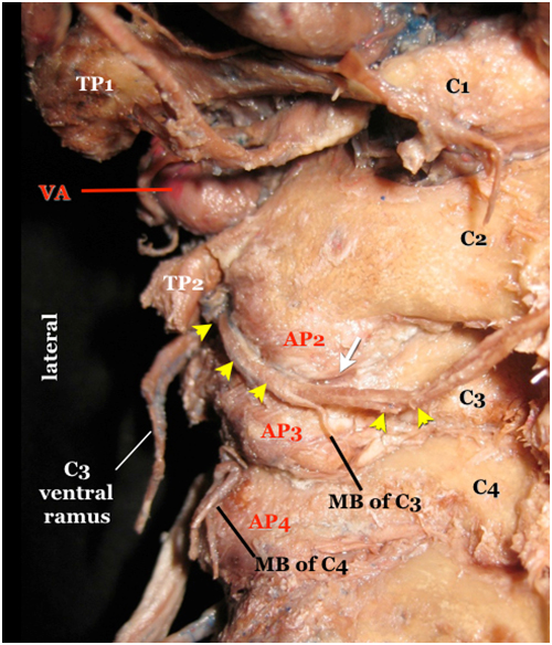
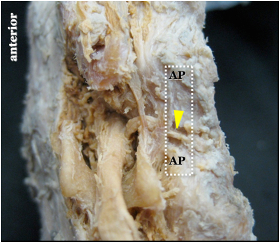
In Figure 1, note that the TON (arrowheads) first appears caudad to the transverse process of axis (TP2) before reaching the surface of the C2/C3 facet joint gap over its lateral aspect. The white arrow indicates the posterior C2/C3 joint gap.

Figure 1 shows a number of important internal anatomical landmarks for localization of the third occipital nerve.

They are:

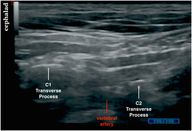
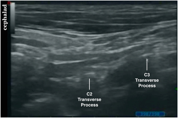
  1. the C1 and C2 transverse processes (bony landmarks);
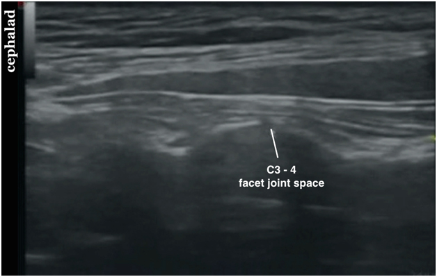
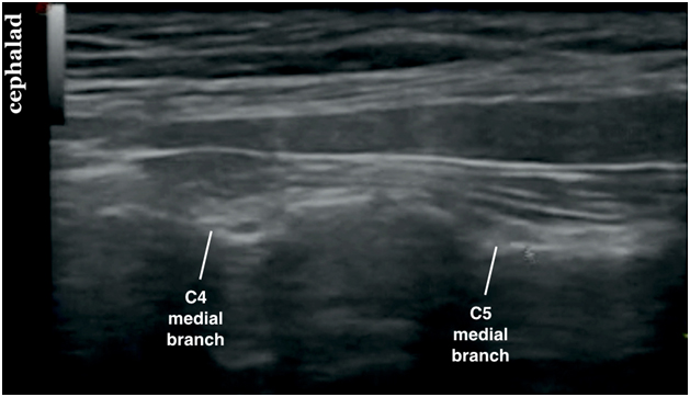
  2. the typical wavy appearance of the lateral aspects of articular pillars of cervical vertebrae (bony landmarks; see ultrasound image); and
  3. the vertebral artery (a vascular landmark) in between the C1/C2 transverse processes.
  4. The Cervical Medial Branches (C-MB)

Figure 1. A posterior view of the cervical spine showing the third occipital nerve in a cadaver specimen

arrowheads = TON; first appears caudad to the transverse process of axis (TP2) before reaching the surface of the C2/C3 facet joint gap over its lateral aspect.
AP = articular process
C1 = atlas, posterior arch
C2 = axis, lamina
MB = medial branch
VA = a segment of the vertebral artery appearing between the transverse process of axis (TP2) and the transverse process of atlas (TP1)
white arrow = the posterior C2/C3 joint gap

Movie 1. TON and cervical medial branch anatomy


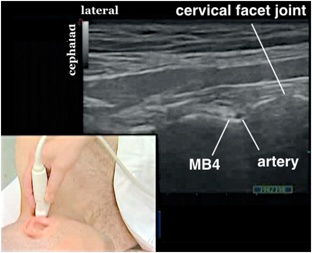
The deep medial branch of C3 dorsal ramus and medial branches of C4-C7 dorsal rami innervate the cervical facet joints. It is important to realize that each facet joint from C3/C4 and below receives innervation from one medial branch above and one below.

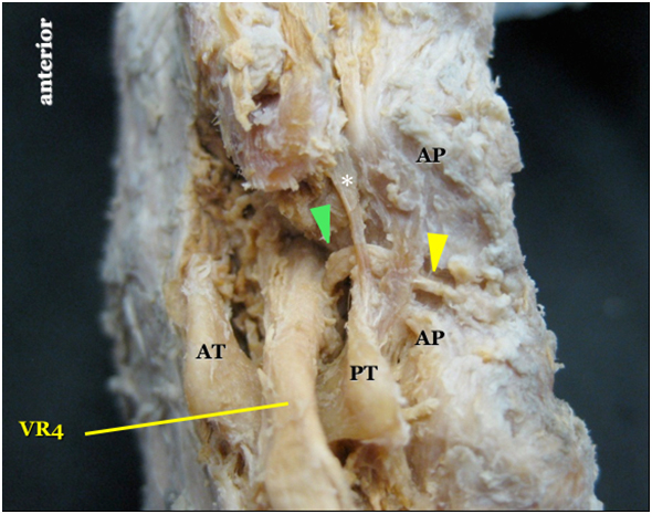
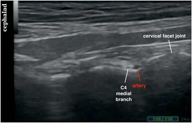
The cervical medial branches are smaller in caliber than the TON. The medial branches emerge from the respective dorsal ramus of a spinal nerve, and each dorsal ramus is held in place by the tendinous parts of the erector spinae muscles. Each cervical medial branch then courses around the waist between the articular processes. For example, the medial branch of C4 is seen within the waist of the superior and inferior articular processes of C4 (Figure 2). The medial branches get progressively smaller at lower cervical levels.

Note that the C4 medial branch (yellow arrowhead in Figure 2) arising from the C4 dorsal ramus (green arrowhead) is held in place by the tendon slips (asterisk) of the erector spinae muscles. The C4 medial branch is located within the waist of the articular process (AP) of C4 cervical vertebra.

Figure 2. Cadaver dissection showing the C4 medial branch

yellow arrowhead = C4 medial branch
green arrowhead = C4 dorsal ramus
* = tendon slip of the erector spinae muscle
AP = articular processes of C4
AT = anterior tubercle of C4 transverse process
PT = posterior tubercle of C4 transverse process
VR4 = the C4 ventral ramus

Movie 2. Cervical medial branch anatomy

Sonoanatomy

The Third Occipital Nerve

Sonographically, the TON appears hypoechoic with internal architecture with an outer hyperechoic border (Figure 3). The nerve lies above the C2-C3 facet joint but it is important to note that the facet joint gap is not regularly seen.

Figure 3. Sonogram showing the third occipital nerve (TON)

TON = third occipital nerve

The Cervical Medial Branches (C-MB)

Sonographically, the medial branches of C4-C7 are also likely hypoechoic but without any obvious internal architecture (Figure 4). They also have a hyperechoic outer border.

Figure 4. Sonogram showing C4 medial branch (MB4)

MB4 = C4 medial branch

Scanning Technique

Position the patient lateral decubitus with the head in the neutral position supported by a pillow.

Use the highest frequency transducer whenever possible since the TON is small in caliber. Imaging a patient with a high BMI can be challenging when a lower frequency transducer is required.

After skin and transducer preparation, place a linear 38-mm high frequency 15 or higher MHz transducer on the skin surface to obtain the best possible transverse view of the TON (Figure 5). It is often necessary to use a 18 MHz transducer to visualize the other smaller medial branches (C4-C7).

Optimize machine imaging capability by selecting the appropriate depth of field, focus range and gain.

Figure 5. A linear transducer positioned longitudinally on the side of the neck to be blocked


The TON is a characteristically hypoechoic structure visualized on top of the C2-C3 facet joint (see US-image in Figure 3).

Figure 6 shows the optimal transducer position to visualize the medial branches (white dotted rectangle).

Figure 6. Optimal transducer (white rectangle) position shown on a cadaver specimen

AP = articular process of facet
white dotted rectangle = optimal transducer position to visualize the medial branches
yellow arrow = medial branch

Nerve Localization

Localization of the Third Occipital Nerve

Palpable external bony landmarks helpful for localizing the TON are:

  1. mastoid process (approximate the C1 vertebra level); and
  2. the angle of the mandible (approximate the C2/C3 vertebra level).

Useful internal sonographic bony landmarks to localize the TON are:

  1. transverse processes of C1-3 and
  2. the C2/C3 facet joint.

First position the transducer longitudinally to locate the mastoid process (MP) (Figure 7).

Figure 7. Sonogram showing the mastoid process

MP = mastoid process

Then move the transducer slightly anteriorly to locate the transverse processes of C1 (atlas) and C2 (axis). The C1 transverse process is significantly bigger and more superficial than C2 (Figure 8). Also, it may be challenging to locate the C2 transverse process that is rudimentary.

Note that the vertebral artery (VA) is located deep between the transverse process of the atlas (TP1) and axis (TP2).

Figure 8. Sonogram showing the C1 and C2 transverse processes


Movie 3. Identifying the mastoid process, C1 and C2 transverse processes


Now move the transducer more caudad to visualize the C3 transverse process (TP3) which is again bigger and more superficial than the C2 transverse process (TP2) (Figure 9).

Figure 9. Sonogram showing the C2 and C3 transverse processes


Then move the transducer posteriorly to capture a longitudinal view of the articular pillars of the C2-C3 facets. The C2/3 facet joint is usually the most cranial facet joint identified in the longitudinal scan. There is an abrupt "drop-off" cranially because of absence of a C1/2 facet joint.

The TON appears hypoechoic with internal architecture with an outer hyperechoic border (Figure 10). The nerve lies above the C2-C3 facet joint but it is important to note that the facet joint gap is not regularly seen.

Figure 10. Sonogram showing the TON above the C2-C3 facet joint

TON = third occipital nerve

Movie 4. Identifying the C3 transverse process and TON


Localization of the Cervical Medial Branches

Then move the transducer slowly caudad to visualize the medial branch of C4 over the groove between the articular processes of C4.

Repeat the same scanning technique more caudad to visualize medial branches of the lower cervical dorsal rami. However, the C5 and distal medial branches may be too small to be visualized.

It is most likely to find medial branches of C4-C7 distinctly hypoechoic with NO internal architecture yet they still have a hyperechoic outer border (Figure 11).

One may visualize small vessels of varying caliber in the vicinity of the cervical medial branches. They are branches of the deep and ascending cervical arteries (Figure 11). Power Doppler may be required to identify these small vessels.

Figure 11. Sonogram showing C4 medial branch


Movie 5. Identifying the cervical medial branches


The peak of the bony outline of the articular processes is where the facet joint lines (CFJ in Figure 12).

Figure 12. Sonogram showing C3-C4 facet joint space


The valley of the articular process contour corresponds to the waist of the articular processes and this is where the medial branches are found (Figure 13).

Figure 13. Sonogram showing 2 consecutive cervical medial branches

Needle Insertion Approach

Ultrasound guided TON and cervical medial branch block is an advanced skill level (level III) block. Identifying the small medial branches over the waists of articular processes requires accurate transducer placement on the articular pillars, high frequency transducer and subtle hand movement. Accurate needle placement is also challenging.

The block is performed with the patient in the lateral decubitus position with the head supported comfortably on a pillow.

The operator is recommended to stand facing the patient with the ultrasound machine behind the patient

We recommend an out-of-plane needle insertion approach with the needle pointing from anterior to posterior (Figure 14). This is important because the needle is pointing away from the vertebral artery which is anterior to the articular processes.

Place a linear 15-18 MHz transducer in the longitudinal plane in the upper neck to capture a long view of the articular processes and facet joints.

Figure 14. Out of plane needle approach to TON

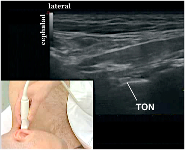
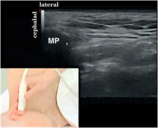
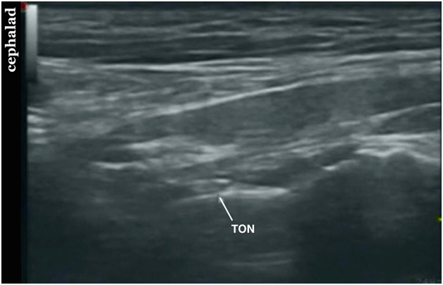
After a systematic anatomical survey and locating the C1, C2 and C3 transverse processes and their corresponding facet joints, identify the TON over the C2/C3 facet joints.

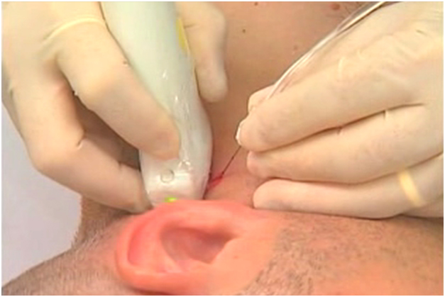
Using the hydrolocation technique with saline, insert a 5 cm short bevel 24 G needle out-of?plane to approach the TON in the anterior to posterior direction (Figure 14).

Aim to see distention of the space surrounding the TON and lifting up of the hyperechoic tissue plane (the tendinous part of the erector spinae muscle) at the time of hydrodissection.

Local Anesthetic Injection

Aim to inject 0.5 mL of local anesthetic around the TON and 0.3 mL for medial branches. The TON and cervical medial branches become more visually apparent after local anesthetic injection. This is especially true with the TON injection.

Movie 6. Out of plane cervical medial branch block and local anesthetic injection


Clinical Pearls

Scanning

It may be challenging to locate the C2 transverse process that is rudimentary. It is best to use the C1 transverse process and the vertebral artery as reference points to locate the TON.

There are hyperechoic lines above the medial branches, These are the tendinous part of erector spinae muscle.

Video Gallery

Selected References

  • Andreas Siegenthaler, Sabine Mlekusch, Sven Trelle, Juerg Schliessbach, Michele Curatolo, and Urs Eichenberger. Accuracy of ultrasound-guided nerve blocks of the cervical zygapophysial joints. 2012;117:347-52.
  • Curatolo M, Eichenberger U. Ultrasound-guided blocks for the treatment of chronic pain. Techniques in Regional Anesthesia and Pain Management 2007;11:95-102.
  • Dash KS, Janis JE, Guyuron B. The lesser and third occipital nerves and migraine headaches. Plast Reconstr Surg 2005;115:1752-8.
  • Eichenberger U, Greher M, Kapral S, Marhofer P. Sonographic visualization and ultrasound-guided block of the third occipital nerve: prospective for a new method to diagnose C2-C3 zygapophysial joint pain. Anesthesiology 2006;104:303-8.
  • Finlayson RJ, Gupta G, Alhujairi M, Dugani S, Tran DQH. Cervical medial branch block: a novel technique using ultrasound guidance. Reg Anesth Pain Med 2012;37:219-23.
  • Finlayson RJ, Etheridge J-PB, Vieira L, Gupta G, Tran DQH. A randomized comparison between ultrasound- and fluoroscopy-guided third occipital nerve block. Reg Anesth Pain Med 2013;38:212-7.
  • Finlayson RJ, Etheridge J-PB, Tiyaprasertkul W, Nelems B, Tran DQH. A prospective validation of biplanar ultrasound imaging for C5-C6 cervical medial branch blocks. Reg Anesth Pain Med 2014;39:160-3.
  • Finlayson RJ, Etheridge J-PB, Tiyaprasertkul W, Nelems B, Tran DQH. A randomized comparison between ultrasound- and fluoroscopy-guided c7 medial branch block. Reg Anesth Pain Med 2015;40:52-7.
  • Kim ED, Kim YH, Park CM, Kwak JA, Moon DE. Ultrasound-guided Pulsed Radiofrequency of the Third Occipital Nerve. Korean J Pain 2013;26:186-90.
  • Lee SH, Kang CH, Lee SH, Derby R, Yang SN, Lee JE, Kim JH, Kim SS, Lee JH. Ultrasound-guided radiofrequency neurotomy in cervical spine: sonoanatomic study of a new technique in cadavers. Clin Radiol 2008;63:1205-12.
  • Narouze S. Ultrasonography in pain medicine: future directions. Techniques in Regional Anesthesia and Pain Management 2009;13:198-202.
  • Narouze S. Atlas of ultrasound-guided procedures in interventional pain management. Springer 2010.
  • Siegenthaler A, Schliessbach J, Curatolo M, Eichenberger U. Ultrasound anatomy of the nerves supplying the cervical zygapophyseal joints: an exploratory study. Reg Anesth Pain Med 2011;36:606-10.
  • Siegenthaler A, Eichenberger U. Ultrasound-guided third occipital nerve and cervical medial branch nerve blocks. Techniques in Regional Anesthesia and Pain Management 2009;13:128-132.
  • Siegenthaler A, Mlekusch S, Trelle S, Schliessbach J, Curatolo M, Eichenberger U. Accuracy of ultrasound-guided nerve blocks of the cervical zygapophysial joints. Anesthesiology 2012;117:347-52.

Share to Facebook Share to Twitter More...