Youtube

Advancing the Science of Ultrasound Guided Regional Anesthesia and Pain Medicine

Transmuscular Quadratus Lumborum Block

Authors

Dr. Jens Børglum
Associate Professor, Consultant, Head of Research
Zealand University Hospital, Copenhagen University
Denmark

Biosketch
Dr. Bernhard Moriggl
Professor, Division of Clinical and Functional Anatomy Department of Anatomy Histology and Embryology
Innsbruck Medical University
Innsbruck, Austria

Biosketch
Dr. Thomas F. Bendtsen
Associate Professor Consultant
Aarhus University Hospital, Aarhus
Denmark

Biosketch

General

Børglum1,2 investigated a new block approach called the transmuscular QL (TQL) block. This block aims to anesthetize branches of the thoracolumbar nerves including the ilioinguinal and iliohypogastric nerves. Preliminary clinical experience suggests that this block can provide pain relief following intra- and retro-peritoneal procedures; e.g., colorectal surgery, laparoscopic nephrectomy, percutaneous nephrolithotomy and laparoscopic cholecystectomy and thoracic procedures such as thoracoscopy and thoracotomy.

When performed bilaterally, this block can control pain following lower abdominal surgery e.g., cesarean section, midline laparotomy and more extensive laparoscopicprocedures e.g., nephrectomy, hemicolectomy, hysterectomy, and bilateral salpingo-oophorectomy.

Preliminary data suggest that TQL block provides predominantly somatic, and to a lesser extent, visceral pain relief, through neural blockade of somatic and sympathetic nerves in the thoracic paravertebral space.

Anatomy

The quadratus lumborum (QL) muscle is a back muscle which attaches via aponeuraotic fibers superiorly to the lower border of the 12th rib, medially to the apex of the transverse processes of L1-L4 through small tendons, and inferiorly to the iliolumbar ligament and the internal lip of the iliac crest (Figure 1).

Action of the quadratus lumborum muscle includes

  1. Lateral flexion of vertebral column, with ipsilateral contraction
  2. Extension of lumbar vertebral column, with bilateral contraction
  3. Could potentially fix the 12th rib during forced expiration
  4. Elevates the ilium (bone), with ipsilateral contraction

Figure 1. Anatomical illustration showing the origin and attachments of the quadratus lumborum muscles.


Other muscular groups in proximity to the QL muscle are (Figure 2):

  1. psoas major (PM) muscle anteriorly,
  2. erector spinae muscle (consisting of the multifidus and the longissimus muscles) posteriorly
  3. anterior abdominal wall muscles (external oblique, internal oblique and transversus abdominis muscles) laterally

Anterior (ventral) to both the QL and psoas major muscles is the peritoneum lining the abdominal cavity containing abdominal visceral organs (Figure 2).

Figure 2. Illustration showing the muscle groups and the surrounding thoracolumbar fascia.


Picture provided by Spine 2006 Lippincott Williams and Wilkins

The muscles are lined by fascial layers. According to Willard3 the so-called two-layer model describes a posterior layer and an anterior layer of the thoracolumbar fascia (TLF) (previously lumbodorsal fascia) (Figure 2). The TLF is a complex set of layers that separates the paraspinal muscles (epaxial muscle compartment) from the muscles of the posterior abdominal wall (hypaxial muscle compartment), i.e. quadratus lumborum (QL) and psoas major (PM).3 The hypaxial and epaxial muscle compartments are separated by an intermuscular septum that medially attaches to the transverse processes of the vertebra. According to the two-layer model this septum is formed by the anterior layer of the TLF (Figure 2).

With the TQL block the anterior layer of the TLF is penetrated with a posterior approach, the needle is advanced further through the QL muscle and finally penetrating anterior layer of the QL investing fascia injecting into the plane between the QL and PM muscles.

The transversalis fascia (TF) is another important fascia in this region. It covers the anterior (ventral) surface of the QL muscle and the antero-lateral surface of the psoas muscle (PM). The TF also covers the anterior (ventral) surface of the transversus abdominis muscle separating this muscle from the peritoneum. At the level of the diaphragm, the TF splits into two layers; one becomes the inferior diaphragmatic fascia and the other courses posterior to the lateral and medial arcuate ligaments and continues with the thoracic endothoracic fascia.

The current hypothesis is that a large volume local anesthetic bolusinjection (e.g., 30 mL) deep to the TF between the QL and PM musclesin the lumbar region will spread cepahlad to reach the thoracic paravertebral space. Widespread thoracolumbar anesthesia from T4 to L1 has been reported following a TQL injection (see Clinical Pearl section).

Scanning Technique

  • Position the patient lateral decubitus (Figure 3) with the side to be blocked uppermost and slightly flexed in the hip and knee joint.
  • Place a pillow under the head and a pillow below the lower flank to facilitate visualization of the QL muscle.
  • It is best for the operator to stand behind the patient since a posterior to anterior in plane needle approach is recommended.
  • After skin and transducer preparation, a curved 2-6 MHz transducer is placed transverse over the flank in the posterior axillary line immediately cranial to the iliac crest (Figure 3)
  • The transducer is then moved slightly posterior and angled caudad until the QL muscle is visualized at the level of the L4 transverse process.
  • The psoas major muscle (PM) is also identified anteriorly and the paraspinal muscle (erector spinae) posteriorly (Figure 3)

Figure 3. Scanning for the TQL block. Patient in the lateral position.

  • The so-called "Shamrock" sign is identified; the L4 transverse process being the stem and the 3 muscles (QL, PM and erector spinae) being the 3 clovers (Figure 4)

Figure 4. A sonogram showing the Shamrock sign.


The stem of the clover is the transverse process of the L4 vertebral body. The three leaves of the clover are the psoas muscle (PM), the quadratus lumborum (QL) muscle attached to the apex of the transverse process, and the erector spinae (ES) muscles.

Anatomical Correlation

Below is a sonogram captured with the subject lying lateral decubitus and a corresponding anatomical dissection (Figure 5). The "Shamrock" sign is clearly illustrated.

Figure 5. Sonogram and anatomical dissection correlation.

PM = Psoas muscle
PSM = Paraspinal muscle
QL = Quadratus lumborum muscle
TP = L4 transverse process
VB = Vertebral body

Here is another anatomical correlation showing ultrasound and an accompanying MRI image of the corresponding region; i.e. ultrasound / MR image fusion. The bony and muscular landmarks are labelled and the thoracolumbar fascia is highlighted in yellow. (Figure 6)

Figure 6. Anatomical correlation between ultrasound and MRI images.

1 and 3 = ultrasound images
2 and 4 = MR images
Yellow line = the thoracolumbar fascia (TLF) as a double line representing the so-called two-layer model described by Willard.3 The posterior layer posterior to the erector spinae muscles and the anterior layer separating the QL muscle and the LG muscle.
White line = the peritoneal recess separating the abdominal wall muscles from the PM.
L4 = vertebral body of L4
LG = longissimus muscle (part of erector spinae muscles)
MF = multifidus muscle (part of erector spinae muscles)
PM = psoas muscle
QL = quadratus lumborum muscle
TP = transverse process

Nerve Localization

The TQL block utilizes the easily recognizable bony landmarks of the vertebral body of L4 and its transverse process as has been previously described with the Shamrock sign.4 The thoracolumbar nerves are normally not visualized but the subcostal nerve (Th12), the ilioinguinal and iliohypogastric nerves (L1) are expected to travel between the anterior investing transversalis fascia (TF) and the QL muscle in an anterolateral direction.

Important internal sonographic landmarks to recognize for the TQL block are as follows:

  1. bone = L4 transverse process and vertebral body
  2. muscles = psoas major muscle (anterior), quadratus lumborum muscle, erector spinae muscles and the anterior abdominal wall muscles (three layers = obliquus externus and internus, transversus abdominis)
  3. peritoneal cavity and abdominal viscera

With the curved 2-6 MHz transducer, it is easy to recognize the "Shamrock" sign - the L4 transverse process (stem) and the 3 muscles, QL muscle, PM muscle anteriorly and erector spinae muscle posteriorly (clover leaves) (Figure 4,5, 6).

Needle Insertion Approach

Position the patient lateral decubitus position with the block side uppermost (Figure 7).

The operator is standing behind the patient.

Place a curved array 2-6 MHz transducer transverse in the posterior axillary line immediately cranial to the iliac crest (Figure 7).

Angle the transducer slightly posterior to identify the Shamrock sign consisting of the QL muscle, PM muscle, erector spinae muscles and the L4 transverse process.

Insert a 100 mm, 21G non-stimulating block needle in-plane from the posterior edge of the transducer in the postero-medial to antero-lateral direction. (Figure 7).

Figure 7. In-plane needle approach for the TQL block.

PM = Psoas major muscle
QL = Quadratus lumborum muscle
ES = Erector spinae muscles
TP = Transverse process of L4

Under ultrasound guidance, advance the needle through the posterior lamina of the thoracolumbar fascia (TLF), the belly of the QL muscle, the anterior layer of the investing QL muscle fascia (Figure 2) until the needle tip has reached the fascial plane between the QL and the PM muscles.

Use hydrodissection and hydrolocation to help identify the needle tip location during needle advancement.

A "pop or click" can be felt and seen when the needle tip penetrates the anterior (ventral) investing QL fascia ventral to the QL muscle.

The optimal final needle position is a point at some distance away (lateral) from the tip of the transverse process. Injection immediately next to the transverse process may accidentally lead to local anesthetic spread towards the lumbar plexus situated within the psoas muscle.

Note that the peritoneal cavity extends deep to the anterior abdominal muscles towards the lateral edge of the QL muscle (Figure 8). Beware of the peritoneal recess that separates the psoas muscle from the abdominal wall muscles.2 The posteromedial to anterolateral needle approach is recommended to avoid accidental puncture of the peritoneum.

Figure 8. The peritoneal cavity extends far laterally and posteriorly deep to the anterior abdominal wall muscles towards the lateral edge of the QL muscle. (Reference: Hansen et al.)2

ESM = Erector spinae muscle
L4 = Vertebral body of L4
PC = Peritoneal cavity
QL = Quadratus lumborum muscle
TP = Transverse process of L4

Local Anesthetic Injection

The TQL block is a large volume block. Care should be taken to dilute the local anesthetic solution if necessary in order to uphold the internationally accepted limits for single bolus injections.5 This is particularly important with bilateral TQL blocks.

The goal is to deposit local anesthetic in the fascial plane between the QL and the PM muscles, deep to the transversalis fascia (Figure 8).

For unilateral block a bolus injection of 30 mL of ropivacaine 0.75% can provide extensive cutaneous anesthesia / analgesia from T4(6) to L1 levels. For bilateral blocks, suggest to administer a total of 60 mL of ropivacaine 0.375%, 30 mL per side. Note to limit the total local anesthetic dose to the maximum recommended dose range.5

Observe the local anesthetic bolus pushing the PM muscle away from the QL muscle and separate the PM muscle from the QL muscle during bolus injection (video).

Aim to promote cranial local anesthetic spread between the 2 muscles. Cephalad spread is required to achieve spread into the thoracic paravertebral space via the pathway posterior to the lateral and medial arcuate ligaments as previously reported by Saito.6

Optimal cephalad local anesthetic spread can be confirmed immediately following block administration by turning the curvilinear array away from the transverse position 90 degrees into the longitudinal position (Figure 9).

Block onset time varies from 10 to 30 minutes after injection.

If the anatomical structures are difficult to distinguish from each other hydrodissection with saline in the fascial plane between the QL and PM muscles are recommended.

Figure 9. Cephalad spread of injected local anesthetic observed with the transducer turned 90 degrees away from the transverse axial position into the longitudinal (parasagittal) transducer position.14

IC = Iliac crest
LA = Local anesthetic
QL = Quadratus lumborum muscle
PM = Psoas muscle

Clinical Pearls

Nerve (Target) Localization

If the L4 transverse process is difficult to visualize it is recommended to focus on the transversus abdominis muscle. Follow the course of the muscle more posterolatteral until it becomes aponeurotic. This aponeurotic structure will normally attach to the posterior side of the QL muscle (Figures 5 and 6) and thus blend with the anterior layer of the thoracolumbar fascia (according to the two-layer model).2

Local Anesthetic Injection

Optimal cranial local anesthetic spread can be monitored and confirmed by turning the transducer 90 degrees away from the transverse axial position to the longitudinal (parasagittal) position to visualize separation of the QL and PM muscles and local anesthetic spread from the iliac crest towards the ribs and the diaphragm (Figure 9).

Local Anesthetic Spread

Comparison with TAP Block

Many transversus abdominis plane (TAP) block techniques, ultrasound or landmark based, have been described over the years. Their analgesic effect, however, may not be consistent. The site and technique of TAP block has been found to influence the extent and pattern of local anesthetic spread.

In a MRI study by Carney,7 subcostal and mid axillary TAP blocks performed anteriorly resulted in contrast local anesthetic spread predominantly in the TAPs of the upper and lower abdominal wall with minimal posterior spread. Similar results were confirmed by Børglum.8 On the other hand, posterior TAP block approaches, i.e., posterior to the mid axillary line and next to the antero-lateral border of the quadratus lumborum (QL) muscle, result in local anesthetic spread not only posteriorly around the QL muscle but far medially to the paravertebral spaces extending from the T5 to L1 vertebral levels.7

Blanco originally described ultrasound-guided (USG) TAP block using a "no-pops" technique in 2007.9 This is the first quadratus lumborum block (QLB1) which aims to deposit local anesthetic at the anterolateral aspect of the QL muscle (Figure 10). The site of needle insertion is very similar to the landmark based Triangle of Petit approach described by McDonnell; i.e. posterior to the mid axillary line.10 Apparently, it also results in a similar spread pattern.7

Figure 10. Schematic diagram of Blanco's quadratus lumborum block (QLB) 1 and 2. (Reference: Blanco et al.)11


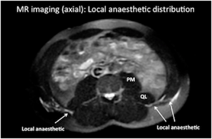
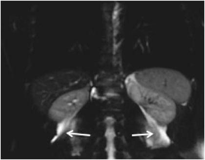
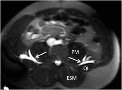
A subsequent study by Børglum demonstrated redundant local anesthetic spread with the QLB1; i.e. lateral and a spread away from the site of injection (Figure 11).12

Figure 11. MRI scan showing dye and local anesthetic spread laterally and anteriorly after a QLB1 injection (Reference: Børglum et al)12

PM = Psoas muscle
QL = Quadratus lumborum muscle

Blanco later abandoned the original QLB1 technique and described the QLB2 which aims to deposit local anesthetic at the posterior aspect of the QL muscle between the QL and the latissimus dorsi muscles.11,13

In contrast with QLB1 and QLB2 blocks, the TQL block aims to inject local anesthetic deep to the transversalis fascia (TF) with a posterior approach into the interfascial space between the QL and psoas major (PM) muscles and contain the local anesthetic spread around and anterior to the QL muscle. Local anesthetic injected anterior to the QL muscle was found to promote cephalad spread within the fascial plane between the QL muscle and PM muscle to the thoracic paravertebral spaces (Figure 12). The spread pattern is posterior to the transversalis fascia (Figure 13) and less "redundant" spread laterally as seen with the original QLB1 injection.

Figure 12. MRI coronal plane view showing cranial dye spread into the thoracic paravertebral space following the bilateral TQL injection (most clearly visualized on the left side)

Arrow = Local anesthetic cephalad spread from the site of lumbar administration at the level of L4.

Figure 13. MRI axial plane view showing dye spread between the psoas major (PM) and QL muscles following a bilateral transmuscular quadratus lumborum block (TQL) injection. The local anesthetic lies deep to the transversalis fascia. ESM = erector spinae muscles.


Mechanism of Action (Anatomical Study)

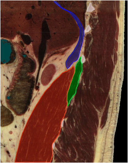
Below is a parasagittal view of the QL muscle, the psoas muscle and the diaphragm (Figure 14). Note that the cephalad parts of the QL muscle and the PM next to the diaphragm create a shape of a funnel which enables the injected local anesthetic trapped in the fascial plane between the two muscle to spread further into the thoracic paravertebral space below the diaphragm. This spread pattern is in accordance with the previous description by Saito.6

This is possible since the QL and PM muscles insert within the thoracic cage (i.e. 12th rib and vertebral body of T12 and disc, respectively) and since the transversalis fascia is continuous with the endothoracic fascia.

Figure 14. A parasagittal view of the quadratus lumborum muscle (green), the psoas muscle (PM) (red) and the diaphragm (blue).

Catheter Insertion

It is technically possible to insert a catheter for continuous TQL block. After needle insertion and generous hydro-dissection with saline to expand the fascial plane between the QL and the PM muscles, a catheter can be advanced 2-3 cm beyond the tip of the needle into the fascial plane. Proper catheter tip position can be confirmed by visualization of appropriate local anesthetic spread during catheter injection.

Total 24 hour dosage of ropivacaine is limited to 800 mg for a grown adult.5

For example, if the initial (bilateral) bolus constitutes 2 x 30 mL ropivacaine 0.375% = 225 mg, it is recommended that the subsequent boluses be2 x 30 mL ropivacaine 0.2% = 120 mg every 12 hours. Thus, the total dosage for 24 hours would be 465 mg.

Repeated boluses twice a day are recommended since the TQL block seems to require a large bolus injection to ensure sufficient cephalad spread into the thoracic paravertebral space.

Video Gallery

The needle penetrates the QL muscle at the posterolateral border with the target endpoint being the fascial plane between the QL muscle and the PM. Try to aim away from the tip of the transverse process as the local anesthetic might then spread along the transverse process toward the lumbar plexus within the psoas muscle. Observe how the two muscles separate from each other.

Selected References

  1. Børglum J, Moriggl B, Jensen K, Lonnqvist PA, Christensen AF, Sauter A, Bendtsen TF. Ultrasound-guided Transmuscular Quadratus Lumborum Blockade. Br J Anaesth 2013, Published 25 March 2013, Online ISSN 1471-6771 - Print ISSN 0007-0912 (http://bja.oxfordjournals.org/forum/topic/brjana_el%3b9919)
  2. Hansen CK, Dam M, Bendtsen TF, Børglum J. Ultrasound-guided quadratus lumborum blocks: Definition of the clinical relevant endpoint of injection and the safest approach. A A Case Rep. 2016; 6(2): 39
  3. Willard FH, Vleeming A, Schuenke MD, Danneels L, Schleip R. The thoracolumbar fascia: anatomy, function and clinical comsiderations. J Anat 2012; 221: 507-36
  4. Sauter AR, Ullensvang K, Niemi G, Lorentzen HT, Bendtsen TF, Børglum J, Pripp AR, Romundstad L. The Shamrock lumbar plexus block: A dose-finding study. Eur J Anaesthesiol 2015; 32: 764-70
  5. Rosenberg PH, Veering BT, Urmey WF. Maximum recommended doses of local anesthetics: a multifactorial concept. Reg Anesth Pain Med 2004;29:564-75
  6. Saito T, Den S, Tanuma K, Tanuma Y, Carney E, Carlsson C. Anatomical bases for paravertebral anesthetic block: fluid communication between the thoracic and lumbar paravertebral regions. Surg Radiol Anat 1999; 21: 359-63
  7. Carney J, Finnerty O, Rauf J, Bergin D, Laffey JG, Mc Donnell JG. Studies on the spread of local anaesthetic solution in transversus abdominis plane blocks. Anaesthesia 2011; 66: 1023-30
  8. Børglum J, Jensen K, Christensen AF, Hoegberg LCG, Johansen SS, Lonnqvist P.-A., Jansen T. Distribution Patterns, Dermatomal Anesthesia, and Ropivacaine Serum Concentrations After Bilateral Dual Transversus Abdominis Plane Block. Reg Anesth Pain Med 2012; 37: 294-301
  9. Blanco R. Tap block under ultrasound guidance: the description of a "no pops" technique. Reg Anesth Pain Med 2007; 32: 130
  10. McDonnell JG, O'Donnell B, Curley G, Heffernan A, Power C, Laffey JG. The analgesic efficacy of transversus abdominis plane block after abdominal surgery: a prospective randomized controlled trial. Anesth Analg 2007; 104: 193-197
  11. Blanco R, Ansari T, Girgis E. Quadratus lumborum block for postoperativ pain after caesarean section: A randomised controlled trial. Eur J Anaesthesiol. 2015 Nov;32(11):812-8
  12. Børglum J, Christensen AF, Hoegberg LCG, Johansen SS, Christensen H, Worm BS, Danker J, Lenz K, Jensen K. Bilateral-dual transversus abdominus (BD-TAP) block or thoracic paravertebral block (TPVB)? Distribution patterns, dermatomal anaesthesia and LA pharmacokinetics. Reg Anesth Pain Med 2012; 37: E136-E139
  13. Blanco R. Optimal point of injection: The quadratus lumborum type I and II blocks (http://www.respond2articles.com/ANA/forums/post/1550.aspx)
  14. Mænchen N, Hansen CK, Dam M, Børglum J. Ultrasound-guided Transmuscular Quadratus Lumborum (TQL) Block for Pain Management after Caesarean Section. Int J Anesthetic Anesthesiol 2016; 3: 048
  15. Dam M, Moriggl B, Hansen CK, Hoermann R, Bendtsen TF, Børglum J. The Pathway of Injectate Spread With the Transmuscular Quadratus Lumborum Block: A Cadaver Study. Anesth Analg 2017 Marr 8. doi: 10.1213/ANE.0000000000001922. [Epub ahead of print]
  16. Dam M, Hansen CK, Børglum J, Chan V, Bendtsen TF. A transverse oblique approach to the transmuscular Quadratus Lumborum block. Anaesthesia 2016;71:603-4

Share to Facebook Share to Twitter More...