Youtube

Advancing the Science of Ultrasound Guided Regional Anesthesia and Pain Medicine

Transversalis Fascia Plane Block

Anatomy

The transversalis fascia plane block, or TFP block, is a truncal block that targets the L1 nerve branches, namely the ilioinguinal and iliohypogastric nerves.The ilioinguinal and iliohypogastric nerves emerge from the lateral border of psoas major muscle, inferior to the 12th rib, and course over the anterior surface of the quadratus lumborum muscle. Lateral to the quadratus lumborum muscle, they initially run deep to transversus abdominis muscle for a variable distance before piercing the transversus abdominis muscle to enter the transversus abdominis plane between the internal oblique and transversus abdominis muscles.

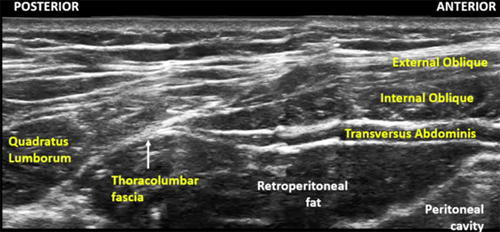
The TFP block targets the ilioinguinal and iliohypogastric nerves where they are between the fascia of the transversus abdominis muscle and the transversalis fascia. The fascia of the transversus abdominis muscle, also called the thoracolumbar fascia, is formed when the transversus abdominis and internal oblique muscles taper off posteriorly into a common aponeurosis and abuts the lateral border of quadratus lumborum muscle. The transversalis fascia is a thin aponeurotic membrane which lies between the transversus abdominis muscle and the extraperitoneal fascia, and is part of the general layer of fascia lining the abdominal cavity.

Anatomical Relationship of the Iliohypogastric and Ilioinguinal Nerves with the Surrounding Muscles


Anterior to the iliac crest, the nerves travel towards the anterior abdominal wall, piercing first the internal oblique muscle, and then the external oblique muscle. However the location at which this occurs varies widely between individuals.

The TFP block is indicated for pain relief following anterior iliac crest bone graft harvesting as the block is performed proximal to the L1 branches that innervate the anterior iliac crest. Local anesthetic spread can also involve the subcostal nerve (T12 spinal nerve). This block is also an analgesic option for inguinal hernia repair, open appendectomy and any surgery involving the L1 dermatome.

The TFP block is different from the transversus abdominis plane (TAP) block in a number of ways. The TFP block is designed to block the L1 nerve branches, which the TAP block does not reliably cover. The TFP block does not cover the dermatomes above L1 and T12 while the TAP block does. Local anesthetic is injected deep to the transversus abdominis muscle for the TFP block versus superficial to the muscle for the TAP block. The site of TFP injection is posterior to the mid-axillary line, unlike the classic ultrasound-guided TAP block.

Target Location of the Transversalis Fascia Plane Block


Scanning Technique

  • Position the patient lateral decubitus and facing the operator with the operative side uppermost. The machine is placed on the opposite side of the bed.
 
  • It is also possible to perform the block with the patient in a supine position.
  • Expose the trunk in the low thoracic and abdominal regions over the axillary line.
  • After skin and transducer preparation, place a curved transducer (2-5 MHz) over the lateral torso just above the iliac crest in the mid axillary line. A low-frequency curved transducer is generappy preferred for its wider field of view and better penetration. If the patient is very slim, a high frequency linear transducer (10-12 MHz) may be used instead.
  • Optimize machine imaging capability; select appropriate depth of field (usually within 5-6 cm), focus range and gain.

Anatomical Correlation

  • Starting anteriorly, the three layers of the abdominal wall can be readily identified. The internal oblique muscle is usually the thickest layer while the transversus abdominis muscle is thinnest and darkest.
  • More posteriorly, the transversus abdominis and internal oblique muscles taper off into their common aponeurosis, the thoracolumbar fascia, and abut against the quadratus lumborum muscle.
 
  • At this point the nerves are sandwiched between the fascia of transversus abdominis and the transversalis fascia.
  • The transversalis fascia is a thin aponeurotic membrane which lies between the transversus abdominis and the extraperitoneal fascia, and is part of the general layer of fascia lining the abdominal cavity.
Schematic Diagram Showing the Muscular Layers, Thoracolumbar Fascia and Transversalis Fascia
 
  • Note that the transversus abdominis and internal oblique taper off posteriorly into a common aponeurosis, also called the thoracolumbar fascia, where they meet the lateral border of quadratus lumborum. The tapered end of transversus abdominis is an important landmark for performing the block.
  • The dark hypoechoic area deep to the tapering off of transversus abdominis is retroperitoneal fat and not the peritoneal cavity. This eliminates concerns of visceral perforation.
  • The target for needle tip placement is just deep to the fascia of transversus abdominis muscle.

Sonogram Showing the Muscular and Fascial Layers Relevant for TFP Block.

Fascial Plane Localization

  • Perform a systematic anatomical survey from medial to lateral and superficial to deep.
  • Identify the external oblique, internal oblique, and transversus abdominis muscles and trace posteriorly until the transversus abdominis muscle tapers off into its aponeurosis, the thoracolumbar fascia.
  • The quadratus lumborum muscle is often visible just posterior to the aponeurosis of the transversus abdominis muscle.
  • Tilting the probe slightly caudad into the pelvis often improves the view of the tapered end of transversus abdominis.

Needle Insertion Approach

In Plane Approach

  • Ultrasound guided TFP block is considered a BASIC skill level block.
  • Insert a 8 cm 22 G insulated block needle in plane to the transducer in an anterior to posterior direction. An echogenic needle is useful in improving visualization of the needle tip given the relatively steep angle and depth.
Picture Showing In Plane Needle Insertion in the Anterior to Posterior Direction
 
  • In the TFP block, the nerves can be consistently targeted where they lie deep to the transversus abdominis muscle, before they ascend into the TAP plane and before they give off their lateral cutaneous branches and branches to the iliac crest.
  • The needle is advanced through the external oblique and internal oblique muscles, aiming for the tapered tip of the transversus abdominis muscle.
  • Needle penetration of the fascial layers between the muscles is signalled by both tactile and fascial pops.
Sonogram Showing Echogenic Needle Advancement
Arrows = Needle
 
  • Once the needle tip has pierced the deep fascia of the transversus abdominis muscle, a test injection is performed. Correct needle tip position is signalled by creation of a pocket of local anesthetic between the transversus abdominis fascia and the transversalis fascia. Injection here produces a visible pocket of local anesthetic that pushes the retroperitoneal fat downwards.
 
Sonogram Showing TFP Block Before and After Local Anesthetic Injection.
EO = external oblique muscle
IO = internal oblique muscle
* and LA = local anesthetic
QL = quadrates lumborum muscle
TA = transversus abdominis muscle
 
Sonogram Showing Final Needle Position and Local Anesthetic Injection
Arrows = needle
* = local anesthetic
IO = internal oblique muscle
TA = transversus abdominis muscle
 
  • If this is not seen, the needle tip has been advanced too far into the retroperitoneal fat and should be withdrawn slightly.
  • On the other hand, if the needle tip has not been advanced far enough, injection above the fascia distends the transversus abdominis muscle, a sign that the needle needs to be advanced one fascial pop deeper.
 

Out of Plane Approach

Coming Soon

Local Anesthetic Injection

  • A volume of 20 mL is generally recommended for injection.
  • For analgesic blocks, a dilute long-acting local anesthetic such as 0.25% bupivacaine or 0.5% ropivacaine may be injected.
  • It is recommended that epinephrine be added to the local anesthetic solution to limit plasma concentrations and reduce the risk of systemic toxicity.

Clinical Pearls

  • In general, the TFP block is a safe block.
  • Block failure can occur, particularly if the sonoanatomy and correct plane for injection are not correctly identified.
  • Local anesthetic systemic toxicity is always a risk to consider, particularly if combined with another regional anesthetic technique, such as a brachial plexus block.
  • The risk can be minimized by the use of epinephrine.
  • Peritoneal puncture is unlikely as the peritoneum does not lie under the transversalis fascia plane in most individuals.

Catheter Insertion

Coming Soon

Image Gallery

Coming Soon

Video Gallery

Selected References

  • Hebbard PD. Transversalis fascia plane block, a novel ultrasound-guided abdominal wall nerve block. Can J Anaesth2009;56:618-620.
  • Chin KJ, Chan V, Hebbard P, Tan JS, Harris M, Factor D. Ultrasound-guided transversalis fascia plane block provides analgesia for anterior iliac crest bone graft harvesting. Can J Anaesth 2012;59:122-3.

Share to Facebook Share to Twitter More...