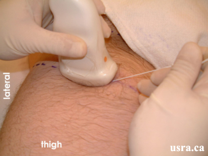
Youtube

Advancing the Science of Ultrasound Guided Regional Anesthesia and Pain Medicine

Sciatic Nerve Block - Proximal Thigh Region

Anatomy

As the sciatic nerve enters the thigh, it courses towards the popliteal fossa behind the femur. At the level of the lesser trochanter, the nerve is often posterior to this bony structure thus access to the sciatic nerve from the anterior thigh can be difficult. The nerve is located on the posterior surface of the adductor magnus muscle within the posterior medial thigh compartment deep to biceps femoris muscle. The sciatic nerve in the thigh is not accompanied by an artery.

The anterior approach to the sciatic nerve targets the nerve in the proximal thigh at the level of the lesser trochanter of the femur.


Transverse View of Proximal Thigh

This figure illustrates the location of the sciatic nerve in the proximal thigh at the level of the lesser trochanter. The white rectangle outlines the path of the ultrasound beam.

Arrowhead = sciatic nerve
1 = adductor magnus muscle
2 = adductor brevis muscle
3 = pectineus muscle
4 = adductor longus muscle
5 = rectus femoris muscle
6 = vastus intermedialis
7 = vastus lateralis
8 = gluteus maximus muscle
F = femur

Scanning Technique

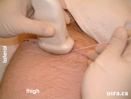
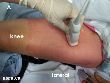
  • Position the patient supine with the hip and knee slightly flexed and the hip externally rotated approximately 45 degrees.
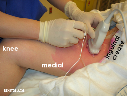
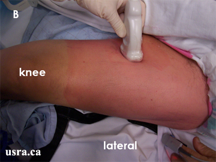
  • After skin and transducer preparation (see transducer preparation section), place a curved low frequency 2-5 MHz transducer firmly on the thigh approximately 8 cm from the inguinal crease.
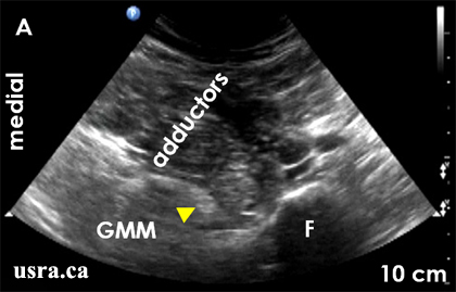
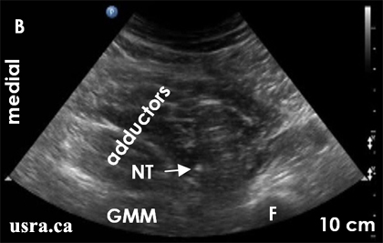
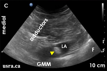
  • Optimize machine imaging capability. Select appropriate depth of field (usually > 6 cm from the skin surface), focus range and gain. The skin to nerve distance is often greater than other sciatic nerve block approaches.
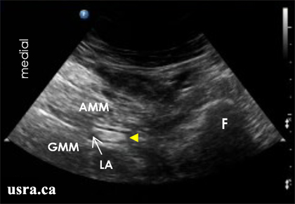
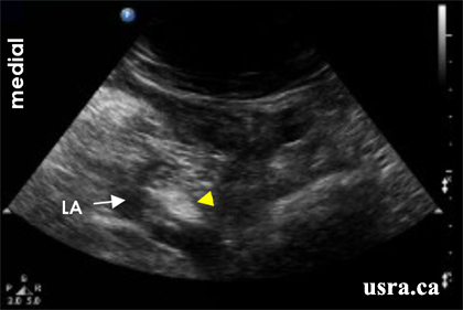
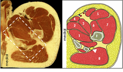
  • Observe on the screen the transverse view of the femur and the sciatic nerve (short axis). The sciatic nerve in the proximal thigh appears predominantly hyperechoic (bright) and often oval or elliptical on ultrasound.
Transducer over the right proximal thigh, approximately 8 cm from the inguinal crease

Anatomical Correlation

Transverse View of Sciatic Nerve in Proximal Thigh

Nerve Localization

  • Perform a systematic anatomical survey from proximal to distal and from lateral to medial.
  • First identify the femur, a curved hyperechoic line with an underlying bone shadow. Move the transducer proximally and distally to identify the lesser trochanter which is often the wide segment immediately above the femoral shaft.
  • Identify the anterior muscular layers: quadriceps muscles laterally and the adductor muscles medially.
  • Identify the gluteus maximus muscle posteriorly. The gluteus maximus muscle bulk gets smaller as the transducer is moved more distally away from the inguinal crease.
  • Locate the hyperechoic sciatic nerve deep to the adductor muscles and posterior to the femur.
  • The sciatic nerve in this region is often elliptical. Visualization can be difficult in the transverse view if the nerve is wide, flat and sandwiched between the adductor and gluteus maximus muscles.
  • If the lesser trochanter obstructs visualization of the sciatic nerve, move the transducer further on the medial aspect of the anterior thigh and orient the transducer in a slightly anterior–posterior direction. This orientation will allow the transducer to capture the best possible transverse view of the sciatic nerve behind (posterior) to the lesser trochanter.
  • Angle the transducer slightly cephalad or caudad to optimize the angle of incidence (90 degrees) to capture the best possible nerve image.
  • Scan the nerve proximally (cephalad) and distally (caudad) to follow the course of the nerve and to confirm nerve identity.

Needle Insertion Approach

  • Ultrasound guided sciatic nerve block in the anterior thigh is considered an INTERMEDIATE to ADVANCED skill level block. Both In Plane (IP) and Out of Plane (OOP) approaches are challenging to perform.
  • The sciatic nerve may be difficult to visualize in this region because of the required depth of beam penetration and the use of a lower frequency transducer. Visualization of the sciatic nerve may be obstructed by the lesser trochanter of the femur and needle tracking can be difficult due to the steep angle of needle trajectory.

In Plane Approach

  • For the In Plane approach, use a 8-12 cm, 22G insulated block needle depending on the thickness of the thigh muscles. Insert the needle on the medial side of the ultrasound transducer after skin local anesthetic infiltration.
  • Advance the needle in a medial to lateral direction as well as an anterior to posterior direction when the thigh is externally rotated.
Advance the needle along the long axis of the transducer in the same plane as the ultrasound beam. This will allow visualization of needle movement in real time.
  • When the thigh is externally rotated, the needle insertion site should be medial to the femoral neurovascular bundle.
  • Unintentional puncture of the femoral vessels is unlikely. The femoral neurovascular bundle is displaced laterally once the thigh is externally rotated.
  • Because of the steep angle of needle advancement, it may be difficult to clearly visualize the block needle. Often, needle and tissue (muscle) movements are observed without a clear view of the needle shaft and tip.
  • Needle to nerve contact is often indicated by nerve movement.
  • Additional confirmation by electrical stimulation is recommended.

Out of Plane Approach

  • For the OOP approach, align the nerve target with the midpoint of the transducer and then insert the block needle in the same location.
  • Clear identification of the needle tip can be technically challenging when the needle angle is steep and the needle is deep inside the muscle layers.
  • Confirm needle to nerve contact by electrical stimulation and observe local anesthetic spread.
Transducer and needle over the right proximal thigh. The thigh is externally rotated. This is an example of the OOP needle approach.

Local Anesthetic Injection

  • Aim to deposit local anesthetic around the sciatic nerve.
  • The sciatic nerve in the proximal thigh is deep thus adjustment in needle direction can be technically challenging. Change in needle angle often requires needle withdrawal and a second needle attempt through the adductor muscles.
  • It is common to find local anesthetic deposited only on one side (the anterior side) of the nerve after injection.
  • Injection to the other side of the nerve may require a separate needle insertion site. A hypoechoic (fluid) expansion can be seen during local anesthetic injection.
  • Fluid expansion in the adductor muscles indicates intramuscular injection. Appropriate needle advancement is required.
  • Inject 15-20 mL of local anesthetic for postoperative analgesia.
Arrowheads = sciatic nerve
AMM = adductor magus muscle
F = femur
GMM = gluteus maximus muscle
LA = local anesthetic around the nerve

Local Anesthetic Spread Around the Sciatic Nerve in the Proximal Thigh

This figure shows further tissue expansion around the sciatic nerve with further local anesthetic injection.

LA = local anesthetic around the nerve

Clinical Pearls

Nerve Localization - Transducer Moved Medially to Image in the Anterior to Posterior Direction

A. The transducer is placed approximately 8 cm from the right inguinal crease. The transducer captures a transverse view in a medial to lateral orientation. In this view, the sciatic nerve image may be obscured by the femur bony shadow.
B. The transducer is now moved more medially and captures a transverse view in the anterior to posterior orientation. The sciatic nerve which is posterior to the femur is more readily visualized in this transducer orientation.

Catheter Insertion

It is uncommon to place a catheter in this location.

Image Gallery

A. Pre Injection
The hyperechoic sciatic nerve (arrowhead) is visualized between the adductor muscles and gluteus maximus muscles (GMM).

F = femur
B. Needle Advancement
The hyperechoic dot representing the needle tip (NT) can be visualized in this case.

F = femur
C. Post Injection
The hyperechoic sciatic nerve (arrowhead) is now surrounded by a collection of hypoechoic local anesthetic (LA).

F = femur
GMM = gluteus maximus muscle

Video Gallery

Selected References

  • Dolan J. Ultrasound-guided anterior sciatic nerve block in the proximal thigh: an in-plane approach improving the needle view and respecting fascial planes. Br J Anaesth 2013;110:319-20.
  • Osaka Y, Kashiwagi M, Nagatsuka Y, Miwa S. Ultrasound-guided medial mid-thigh approach to sciatic nerve block with a patient in a supine position. J Anesth 2011;25:621-4.
  • Moayeri N, van Geffen GJ, Bruhn J, Chan VW, Groen GJ. Correlation among ultrasound, cross-sectional anatomy, and histology of the sciatic nerve: a review. Reg Anesth Pain Med 2010;35:442-9.
  • Latzke D, Marhofer P, Zeitlinger M, Machata A, Neumann F, Lackner E, Kettner SC. Minimal local anaesthetic volumes for sciatic nerve block: evaluation of ED99 in volunteers. Br J Anaesth 2010;104:239-44.
  • Ota J, Sakura S, Hara K, Saito Y. Ultrasound-Guided Anterior Approach to Sciatic Nerve Block: A Comparison with the Posterior Approach. Anesth Analg 2009;108:660-5.
  • Chan VW, Nova H, Abbas S, McCartney CJ, Perlas A, Quan XD: Ultrasound Examination and Localization of the Sciatic Nerve: A Volunteer Study. Anesthesiology 2006; 104: 309-14.

Share to Facebook Share to Twitter More...