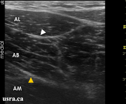
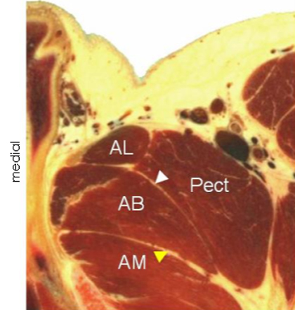
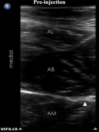
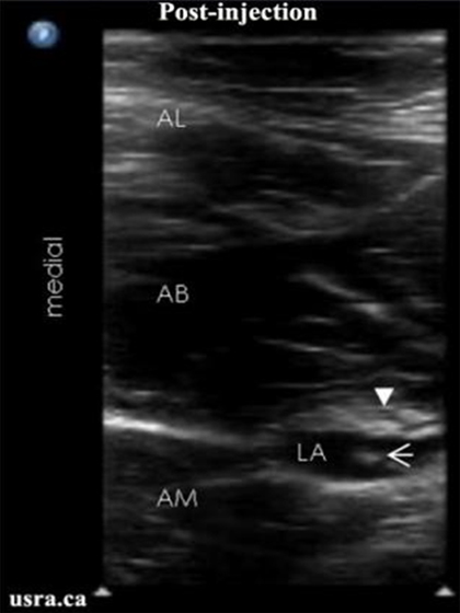
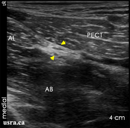
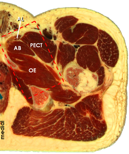
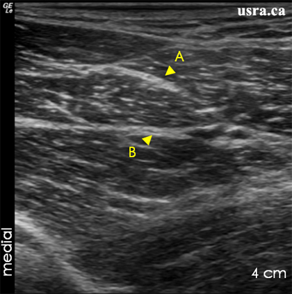
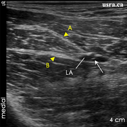
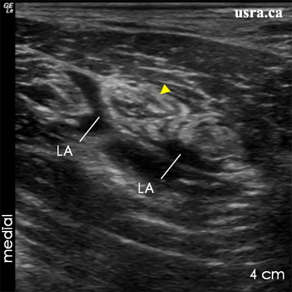
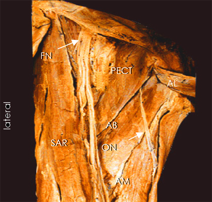
Youtube

Advancing the Science of Ultrasound Guided Regional Anesthesia and Pain Medicine

Obturator Nerve Block

Anatomy

The obturator nerve (L2-4) descends towards the pelvis from the medial border of the psoas major muscle. After passing through the obturator foramen, the obturator nerve enters medial aspect of the thigh, and divides into an anterior branch between the adductor longus and brevis muscles and a posterior branch between the adductor brevis and magnus muscles. The posterior branch usually gives off an articular branch to the knee joint.

The obturator nerve provides sensory innervation to a relatively small area on the medial aspect of the knee. However, blockade of the obturator nerve can decrease opioid requirements and improve analgesia following knee replacement surgery when combined with femoral and sciatic nerve blocks.

AB, AL and AM = adductor brevis, longus and magnus muscles
FN = femoral nerve
ON = obturator nerve
PECT = pectineus muscle
SAR = sartorius muscle

Scanning Technique

  • Position the patient supine with the leg to be blocked slightly externally rotated.
  • Expose the groin and the medial aspect of the proximal thigh.
  • After skin and transducer preparation, place a linear transducer with the appropriate frequency range (10-12 MHz) in the inguinal crease and scan slightly distally in the upper medial thigh.
  • Optimize machine imaging capability; select appropriate depth of field (usually within 2-4 cm), focus range and gain.
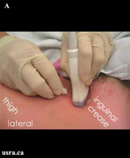
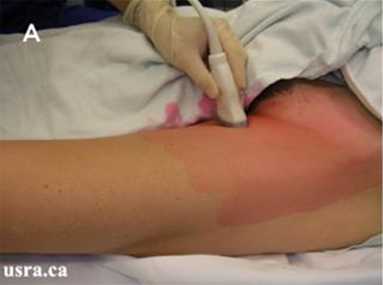
Figure A. A high frequency linear transducer is placed on the medial side of the left thigh below the inguinal crease.
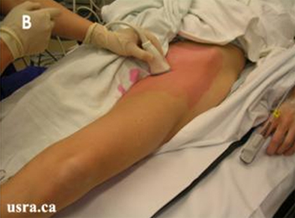
Figure B. Note the left leg is slightly externally rotated.

Anatomical Correlation

The Obturator Nerve in the Proximal Thigh

AB = adductor brevis muscle AL = adductor longus muscle AM = adductor magnus muscle PECT = pectineus muscle
White arrowhead = anterior branch of obturator nerve Yellow arrowhead = posterior branch of obturator nerve

Nerve Localization

  • Perform a systematic anatomical survey on the medial aspect of the proximal thigh.
  • The two branches of the obturator nerve are expected to lie within the fascial planes between the adductor muscles.
  • Identify the femoral vein below the inguinal crease and then move the linear transducer medially.
  • Identify the pectineus and adductor longus muscles located medially. The anterior branch of the obturator nerve (usually a hyperechoic structure) can be found between the adductor longus (more superficial) and brevis muscles.
  • The posterior branch (often hyperechoic) is found between the adductor brevis (more superficial) and magus muscles.
  • Trace the course of the obturator nerve proximally and distally for a short distance.
  • Low current electrical stimulation of the obturator nerve will elicit adductor muscle contraction and aid nerve localization.

Needle Insertion Approach

Ultrasound guided obturator nerve block is considered a INTERMEDIATE skill level block. It can be challenging to image the obturator nerve because of its small size.

In Plane Approach

  • This is a good alternative approach for obturator nerve block.
  • Insert a 8 cm 22 G insulated needle inline with the ultrasound beam so that the needle shaft and tip can be visualized.
  • Again identify the anterior and posterior branches of the obturator nerve before local anesthetic injection.

Out of Plane Approach

  • Insert a 5 cm 22 G insulated needle perpendicular to the transducer and the ultrasound beam (see picture below).
  • Identify the anterior and posterior branches of the obturator nerve lying in the fascial plane between the adductor longus and brevis muscles and the adductor brevis and magnus muscles respectively.
  • Confirm the identity of the branches of the obturator nerve by electrical stimulation and differentiate between the anterior and posterior divisions.
Figure A. Out of plane needle insertion on the medial side of the left leg below the inguinal crease.
Figure B. Needle (arrow) approaching the anterior branch of the obturator nerve (arrowhead).

Local Anesthetic Injection

  • Inject 5-10 mL of local anesthetic in each of the 2 intermuscular fascial planes. Observe local anesthetic distention of the intermuscular planes and local anesthetic surrounding the hyperechoic nerve structures.
  • Scan proximally and distally to assess the extent of local anesthetic spread.

Injection to the Posterior Branch of Obturator Nerve

AB, AL and AM = adductor brevis, longus and magus muscles
Arrowhead = posterior branch of obturator nerve
AB, AL and AM = adductor brevis, longus and magus muscles
Arrow = needle tip
Arrowhead = posterior branch of obturator nerve
LA = local anesthetic

Clinical Pearls

Needle Insertion and Local Anesthetic Injection

  • Visualization of the needle tip with the out of plane approach can be challenging because of the depth required to reach the posterior branch.
  • The "hydro location" technique can be very useful to localize the needle tip.
  • The "hydro dissection" technique is also useful to "dissect" out the intermuscular fascial plane before local anesthetic injection.

Catheter Insertion

Catheter insertion is seldom indicated for this block.

Image Gallery

Pre Injection Scan
The obturator nerve (arrowheads) appears strongly hyperechoic immediately below the inguinal crease.

AB = adductor brevis muscle externus muscle
AL = adductor longus muscle
OE = obturator externus muscle
PECT = pectineus muscle

Anatomical Correlation

Obturator Nerve in the Proximal Thigh Immediately Below the

AB = adductor brevis muscle
AL = adductor longus muscle
OE = obturator externus muscle
PECT = pectineus muscle
Detailed anatomy within the scanned area (red dotted rectangle).
The obturator nerve (arrowheads) is located between the pectineus (PECT) and adductor brevis (AB) muscles.

AL = adductor longus muscle
FV = femoral vein
OE = obturator externus muscle
Pre Injection Scan
As the transducer is moved more distally away from the inguinal crease, the obturator nerve is seen to divide into the anterior (A) and posterior (B) branches.
Post Injection Scan
A small amount of local anesthetic (LA) has been injected through the needle. Note the needle is visualized as a hyperechoic dot (arrow) in cross section.
Post Injection Scan
Local anesthetic (LA) is now visualized around the nerve (arrowhead) and has distended the intermuscular fascial plane.

Video Gallery

Selected References

  • Feigl GC, Ulz H, Pixner T, Dolcet C, Likar R, Sandner-Kiesling A. Anatomical investigation of a new vertical obturator nerve block technique. Ann Anat 2013;195:82-7.
  • Manassero A, Bossolasco M, Ugues S, Palmisano S, De Bonis U, Coletta G. Ultrasound-guided obturator nerve block: interfascial injection versus a neurostimulation-assisted technique. Reg Anesth Pain Med 2012;37:67-71.
  • Paraskeuopoulos T, Saranteas T. Ultrasound-guided obturator nerve block: the importance of the medial circumflex femoral vessels. Reg Anesth Pain Med 2012;37:565-6.
  • Taha AM. Ultrasound-Guided Obturator Nerve Block: A Proximal Interfascial Technique. Anesth Analg 2012;114:236-9.
  • Lee SH, Jeong CW, Lee HJ, Yoon MH, Kim WM. Ultrasound guided obturator nerve block: a single interfascial injection technique. J Anesth 2011;25:923-6.
  • Snaith R, Dolan J. Ultrasound-guided interfascial injection for peripheral obturator nerve block in the thigh. Reg Anesth Pain Med 2010;35:314-5.
  • Akkaya T, Ozturk E, Comert A, Ates Y, Gumus H, Ozturk H, Tekdemir, Elhan A. Ultrasound-Guided Obturator Nerve Block: A Sonoanatomic Study of a New Methodologic Approach. Anesth Analg 2009;108:1037-41.
  • Fujiwara Y, Sato Y, Kitayama M, Shibata Y, Komatsu T, Hirota K. Obturator Nerve Block: From Anatomy to Ultrasound Guidance. Anesth Analg 2008;106:350-1.
  • Saranteas T, Paraskeuopoulos T, Alevizou A, Kouskouri A, Zogojiannis J, Anagnostopoulou S, Chantzi C. Identification of the obturator nerve divisions and subdivisions in the inguinal region: a study with ultrasound. Acta Anaesthesiol Scand 2007;51:1404-1406.
  • Fujiwara Y, Sato Y, Kitayama M, Shibata Y, Komatsu T, Hirota K. Obturator nerve block using ultrasound guidance. Anesth Analg 2007;105:888-889.
  • Helayel P E, da Conceicao D B, Pavei P, Knaesel J A, Oliveira Filho G R. Ultrasound-guided obturator nerve block: a preliminary report of a case series. Reg Anesth Pain Med 2007;32:221-226.
  • Soong J, Schafhalter-Zoppoth I, Gray A T. Sonographic imaging of the obturator nerve for regional block. Reg Anesth Pain Med 2007;32:146-151.

Share to Facebook Share to Twitter More...