Youtube

Advancing the Science of Ultrasound Guided Regional Anesthesia and Pain Medicine

Saphenous Nerve Block

Anatomy

The saphenous nerve is the largest cutaneous branch of the femoral nerve. In the proximal thigh, the saphenous nerve often lies anterior to the femoral artery as this vessel passes beneath the sartorius muscle, and posterior to the aponeurotic covering of the adductor canal. The saphenous nerve descends along the medial side of the knee posterior to the sartorius muscle.


In the distal thigh, the saphenous nerve pierces the fascia lata between the tendons of the sartorius and gracilis muscles (see picture), and then becomes a subcutaneous nerve. The saphenous nerve may also surface between the sartorius and vastus medialis muscles. Below the knee, the nerve passes along the tibial side of the leg, adjacent to the great saphenous vein subcutaneously. At the ankle, one branch of the nerve is located on the medial side next to the subcutaneous saphenous vein.

Scanning Technique

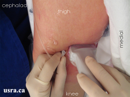
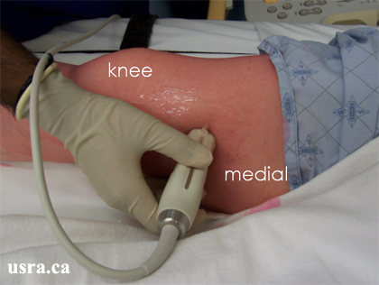
  • Position the patient supine with the leg slightly externally rotated.
  • Expose the lower part of the thigh, knee and upper leg.
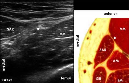
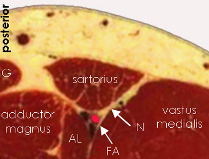
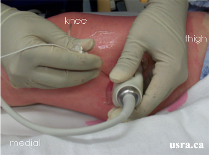
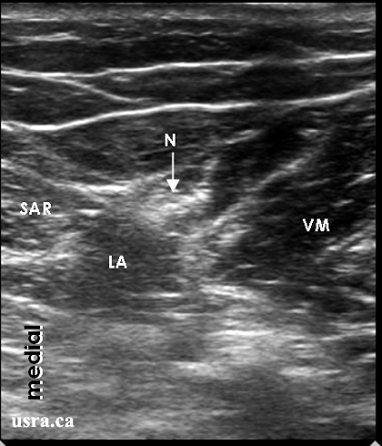
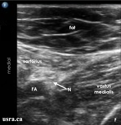
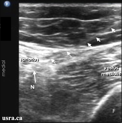
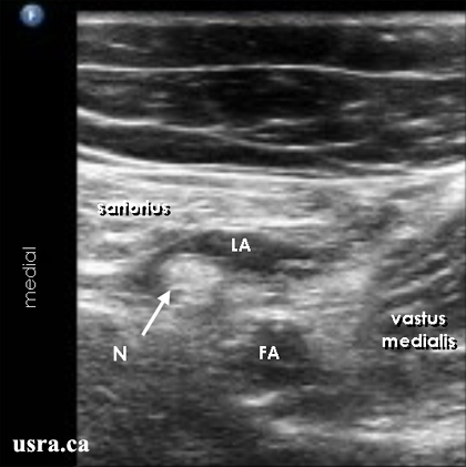
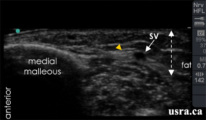
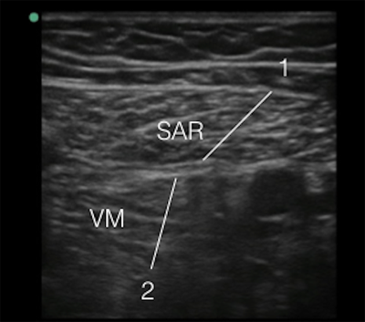
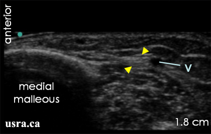
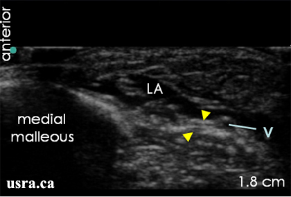
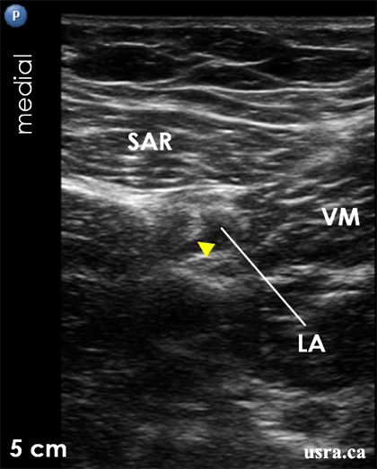
  • After skin and transducer preparation, place a linear transducer with the appropriate frequency range (10-12 MHz) starting in the proximal thigh and scan distally to the knee. The saphenous nerve can be blocked reliably in the distal 1/3 of the thigh.
  • Optimize machine imaging capability; select appropriate depth of field (usually within 1-3 cm), focus range and gain.
Transducer over the medial side of the right thigh.

Anatomical Correlation

Saphenous Nerve in the Distal Thigh Above the Knee

AM = adductor magnus muscle
Arrowhead = saphenous nerve
GR = gracilis muscle
SAR = sartorius muscle
SM = semimembranosus muscle
VM = vastus medialis muscle

Nerve Localization

  • Perform a systematic anatomical survey from proximal thigh to distal thigh. The saphenous nerve is often predominantly hyperechoic. This small nerve is sometimes challenging to visualize in the thigh and leg.
  • Identify the femoral artery and the sartorius muscle. The sartorius muscle is noted to cross the femoral artery as the muscle travels from lateral (anterior superior iliac spine) to medial (superior part of medial surface of tibia).
  • In the distal thigh, the saphenous nerve is often located deep (posterior) to the sartorius muscle in the subsartorial compartment. The nerve lies next to the femoral artery which eventually descends deep into the adductor canal.
  • A motor branch of the femoral nerve supplying the vastus medialis muscle is also found in the subsartorial compartment.
  • Electrical stimulation may be used to differentiate the saphenous nerve (sensory stimulation) and the nerve to the vastus medialis muscle (motor stimulation) in the distal thigh.
  • More distally in the thigh, the saphenous nerve becomes superficial and can be found in the fascial plane between the vastus medialis and the sartorius muscles.
The saphenous nerve (N) is located deep to the sartorius muscle and adjacent to the femoral artery in this case.

AL = adductor longus muscle
FA = femoral artery
G = gracilis muscle
N = saphenous nerve

Needle Insertion Approach

In Plane Approach

  • Ultrasound guided saphenous nerve block is considered a INTERMEDIATE skill level block. It can be challenging to image this small nerve in some individuals.
  • Insert a 5-8 cm 22 G needle parallel to and inline with the transducer and the ultrasound beam.
The transducer is over the medial aspect of the right distal thigh; the block needle is inserted in the lateral to medial direction using the in plane approach.
  • Aim to place the needle into the fascial plane between the sartorius and vastus medialis muscles.
  • Use an insulated needle if electrical stimulation of the nerve (motor branch) to the vastus medialis muscle is intended.

Out of Plane Approach

The OOP approach is also commonly used for saphenous nerve block. The needle tip is more difficult to visualize but the needle to nerve distance is shortest using this approach.

Local Anesthetic Injection

  • If the saphenous nerve is visualized (a predominantly hyperechoic structure), inject 5-10 mL of local anesthetic around the nerve.
  • If the nerve is not clearly visualized in the distal thigh, local anesthetic injection is recommended into the fascial plane between the vastus medialis and sartorius muscles. Injection of an additional 5-10 mL deep to the sartorius muscle is also recommended.
LA = local anesthetic
N = saphenous nerve
SAR = sartorius muscle
VM = vastus medialis muscle
Local Anesthetic Injection for Saphenous Nerve Block in the Distal Thigh.
The saphenous nerve is located between the sartorius and vastus medialis muscles.

F = femur
FA = femoral artery
N = saphenous nerve
Needle Insertion
The needle is inserted from lateral to medial using the in plane approach.

Arrows = block needle
F = femur
N = saphenous nerve
Post Injection
A hypoechoic local anesthetic (LA) collection is seen surrounding the saphenous nerve (N).

FA = femoral artery

Clinical Pearls

Nerve Localization - Saphenous Nerve in Other Locations

The saphenous nerve can be located more distally and subcutaneously in the following locations:

  1. between the sartorius and gracilis muscles in the thigh immediately above the knee
  2. in the medial side of the leg just below the knee at the level of the tibial tubercle where the saphenous nerve lies next to the saphenous vein subcutaneously
  3. in the middle half of the leg where the nerve is adjacent to the subcutaneous saphenous vein
  4. at the level of the ankle where the nerve is next to the subcutaneous saphenous vein
Saphenous nerve at the ankle level.

Arrowhead = saphenous nerve
SV = saphenous vein
  • It can be challenging to identify the saphenous nerve below the knee since it is small and located in subcutaneous tissue. In this case, it may be useful to place a tourniquet around the leg so that the subcutaneous saphenous vein becomes distended and easily visible. The saphenous nerve often lies immediately adjacent to the vein.

Adductor Canal Block

  • The recently termed adductor canal block refers to saphenous nerve block in the subsartorial compartment in the proximal thigh. The adductor canal is marked by the apex of the femoral triangle to the adductor hiatus. Anatomically, the apex of the femoral triangle is defined by the crossing of the medial margin of the adductor longus muscle and the medial margin of the sartorius muscle.

  • The adductor canal contains the femoral vessels, the saphenous nerve, and the nerve to the vastus medialis muscle. It also contains the obturator nerve (the posterior division) which often enters the distal part of the canal and exits through the hiatus of the adductor magnus tendon to the popliteal fossa accompanied by the femoral artery.
  • The advantage of the adductor canal block is sparing of the motor fibers to the quadriceps muscles thus preserving muscle strength following knee surgery and allowing early ambulation and rehabilitation as compared to conventional femoral nerve block. The single shot injection approach, in plane or out of plane, is the same as for saphenous nerve block. The authors generally inject 20 mL of 0.5% ropivacaine for total knee replacement surgery.

Needle Insertion Technique

  • During needle advancement, it is important to penetrate 2 layers of fascia/membrane to enter the adductor canal. The first one is the fascia surrounding the sartorius muscle and the deeper one is the vastoadductor membrane as shown in the figure below.
Adductor Canal Anatomy

1 = fascial lining of sartorius muscle
2 = vastoadductor membrane
SAR = sartorius muscle
VM = vastas medialis muscle

Catheter Insertion

  • A catheter may be inserted for continuous saphenous nerve block but this is not commonly performed.
  • See Catheter Technique for the principles of catheter insertion.
  • Note that the catheter exit site and dressing may be too close to the surgical site when it is inserted prior to total knee replacement surgery. Discuss with the surgeon about the optimal timing and location of catheter insertion.
  • Also note that the catheter exit site is under the thigh tourniquet. Consider the risk of double crush syndrome.

Image Gallery

Saphenous Nerve in the Ankle Region

Pre Injection
The saphenous nerve (arrowheads) is next to the saphenous vein (V). Both structures are superficial in the subcutaneous plane.
Post Injection
A wall of local anesthetic (LA) is visualized in the subcutaneous tissue superficial to the saphenous nerve and vein (V).

Arrowheads = saphenous nerve

Saphenous Nerve in the Distal Thigh

Arrowhead = saphenous nerve
SAR = sartorius muscle
VM = vastus medialis muscle
Arrowhead = saphenous nerve
LA = local anesthetic
SAR = sartorius muscle
VM = vastus medialis muscle

Video Gallery

Selected References

  • Bendtsen TF, Moriggl B, Chan V, Pedersen EM, Borglum J. Defining adductor canal block. Reg Anesth Pain Med 2014;39:253-4.
  • Kim DH, Lin Y, Goytizolo EA, Kahn RL, Maalouf DB, Manohar A, Patt ML, Goon AK, Lee Y-Y, Ma Y, Yadeau JT. Adductor Canal Block versus Femoral Nerve Block for Total Knee Arthroplasty: A Prospective, Randomized, Controlled Trial. Anesthesiology 2014;120:540-50.
  • Damarey B, Demondion X, Wavreille G, Pansini V, Balbi V, Cotten A. Imaging of the nerves of the knee region. Eur J Radiol 2013;82:27-37.
  • Espelund M, Fomsgaard JS, Haraszuk J, Mathiesen O, Dahl JB. Analgesic efficacy of ultrasound-guided adductor canal blockade after arthroscopic anterior cruciate ligament reconstruction: a randomised controlled trial. Eur J Anaesthesiol 2013;30:422-8.
  • Hanson NA, Derby RE, Auyong DB, Salinas FV, Delucca C, Nagy R, Yu Z, Slee AE. Ultrasound-guided adductor canal block for arthroscopic medial meniscectomy: a randomized, double-blind trial. Can J Anaesth 2013;60:874-80.
  • Ishiguro S, Asano N, Yoshida K, Nishimura A, Wakabayashi H, Yokochi A, Hasegawa M, Sudo A, Maruyama K. Day zero ambulation under modified femoral nerve block after minimally invasive surgery for total knee arthroplasty: preliminary report. J Anesth 2013;27:132-4.
  • Jager P, Zaric D, Fomsgaard JS, Hilsted KL, Bjerregaard J, Gyrn J, Mathiesen O, Larsen TK, Dahl JB. Adductor Canal Block Versus Femoral Nerve Block for Analgesia After Total Knee Arthroplasty: A Randomized, Double-blind Study. Reg Anesth Pain Med 2013;38:526-32.
  • Kent ML, Hackworth RJ, Riffenburgh RH, Kaesberg JL, Asseff DC, Lujan E, Corey JM. A Comparison of Ultrasound-Guided and Landmark-Based Approaches to Saphenous Nerve Blockade: A Prospective, Controlled, Blinded, Crossover Trial. Anesth Analg 2013;117:265-70.
  • Kwofie MK, Shastri UD, Gadsden JC, Sinha SK, Abrams JH, Xu D, Salviz EA. The effects of ultrasound-guided adductor canal block versus femoral nerve block on quadriceps strength and fall risk: a blinded, randomized trial of volunteers. Reg Anesth Pain Med 2013;38:321-5.
  • Moore DM, O'Gara A, Duggan M. Continuous Saphenous Nerve Block for Total Knee Arthroplasty: When and How. Reg Anesth Pain Med 2013;38:370-1.
  • Andersen HL, Gyrn J, Moller L, Christensen B, Zaric D. Continuous Saphenous Nerve Block as Supplement to Single-Dose Local Infiltration Analgesia for Postoperative Pain Management After Total Knee Arthroplasty. Reg Anesth Pain Med 2012;38:106-11.
  • Ishiguro S, Yokochi A, Yoshioka K, Asano N, Deguchi A, Iwasaki Y, Sudo A, Maruyama K. Technical communication: anatomy and clinical implications of ultrasound-guided selective femoral nerve block. Anesth Analg 2012;115:1467-70.
  • Lopez AM, Sala-Blanch X, Magaldi M, Poggio D, Asuncion J, Franco CD. Ultrasound-guided ankle block for forefoot surgery: the contribution of the saphenous nerve. Reg Anesth Pain Med 2012;37:554-7.
  • Jaeger P, Grevstad U, Henningsen MH, Gottschau B, Mathiesen O, Dahl JB. Effect of adductor-canal-blockade on established, severe post-operative pain after total knee arthroplasty: a randomised study. Acta Anaesthesiol Scand 2012;56:1013-9.
  • Kapoor R, Adhikary SD, Siefring C, McQuillan PM. The saphenous nerve and its relationship to the nerve to the vastus medialis in and around the adductor canal: an anatomical study. Acta Anaesthesiol Scand 2012;56:365-7.
  • Ishiguro S, Yokochi A, Yoshioka K, Asano N. Anatomy and Clinical Implications of Ultrasound-Guided Selective Femoral Nerve Block. Anesth Anesth 2012;115:1467-70.
  • Lundblad M, Forssblad M, Eksborg S, Lonnqvist P-A. Ultrasound-guided infrapatellar nerve block for anterior cruciate ligament repair: a prospective, randomised, double-blind, placebo-controlled clinical trial. Eur J Anaesthesiol 2011;28:511-8.
  • Saranteas T, Anagnostis G, Paraskeuopoulos T, Koulalis D, Kokkalis Z, Nakou M, Anagnostopoulou S, Kostopanagiotou G. Anatomy and clinical implications of the ultrasound-guided subsartorial saphenous nerve block. Reg Anesth Pain Med 2011;36:399-402.
  • Lund J, Jenstrup MT, Jaeger P, Sorensen AM, Dahl JB. Continuous adductor-canal-blockade for adjuvant post-operative analgesia after major knee surgery: preliminary results. Acta Anaesthesiol Scand 2011;55:336-42.
  • Manickam B, Perlas A, Duggan E, Brull R, Chan VWS, Ramlogan R. Feasibility and efficacy of ultrasound-guided block of the saphenous nerve in the adductor canal. Reg Anesth Pain Med 2009;34:578-80.
  • Horn J-L, Pitsch T, Salinas F. Anatomic basis to the ultrasound-guided approach for saphenous nerve blockade. Reg Anesth Pain Med 2009;34:486-9.
  • Bianchi S, Martinoli C. Ultrasound of the nerves of the knee region: Technique of examination and normal US appearance. J Ultrasound 2007;10: 68-75.
  • Lundblad M, Kapral S, Marhofer P, Lonnqvist PA. Ultrasound-guided infrapatellar nerve block in human volunteers: description of a novel technique. Br J Anaesth 2006;97:710-4.
  • Krombach J, Gray AT: Sonography for saphenous nerve block near the adductor canal. Reg Anesth Pain Med 2007; 32: 369-70
  • Lundblad M, Kapral S, Marhofer P et al. Ultrasound-guided infrapatellar nerve block in human volunteers: description of a novel technique. Br J Anaesth 2006;97: 710-714.
  • Gray A T, Collins A B. Ultrasound-guided saphenous nerve block. Reg Anesth Pain Med 2003;28:148.

Share to Facebook Share to Twitter More...