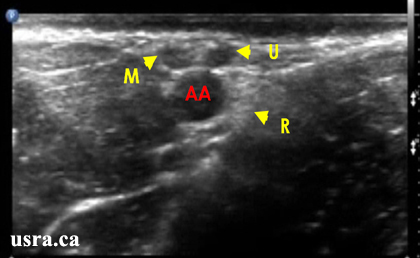
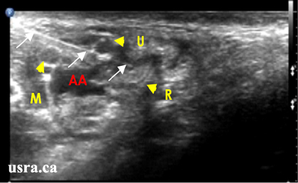
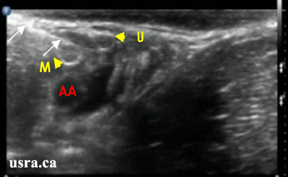
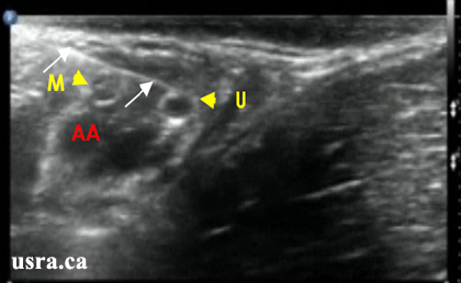
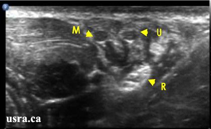
Youtube

Advancing the Science of Ultrasound Guided Regional Anesthesia and Pain Medicine

Axillary Block

Anatomy

The brachial plexus originates from the ventral primary rami of spinal nerves C5-T1 and extends from the neck to the apex of the axilla. The brachial plexus is an intricate network of nerves described from proximal to distal as follows: Roots (interscalene region), Trunks and Divisions (supraclavicular region), Cords (infraclavicular region) and Terminal Branches (axillary region).

In general, the brachial plexus provides sensory and motor innervation to the upper limb although there are a few exceptions: the lateral pectoral nerve (C5-7) and the medial pectoral nerve (C8, T1) supply the pectoral muscles; the long thoracic nerve (C5-7) supplies the serratus anterior muscle; the thoracodorsal nerve (C6-8) supplies the latissimus dorsi muscle and the suprascapular nerve supplies the supraspinatus and infraspinatus muscles.

AXI = axillary nerve
C5-8 = cervical roots 5-8
T1 = first thoracic root
MED = median nerve
MC = musculocutaneous nerve
RAD = radial nerve
ULN = ulnar nerve

The axillary block aims to block the terminal branches of the brachial plexus which include the median, ulnar, radial and musculocutaneous nerves. The musculocutaneous nerve often departs from the lateral cord in the proximal axilla and is commonly spared by the axillary approach. The median, ulnar and radial nerves lie next to the axillary artery and are surrounded by the biceps, coracobrachialis and triceps muscles. It is important to locate and occlude the axillary vein(s) by transducer applied pressure to avoid unintentional intravascular injection.

Transverse view of the Axilia

Scanning Technique

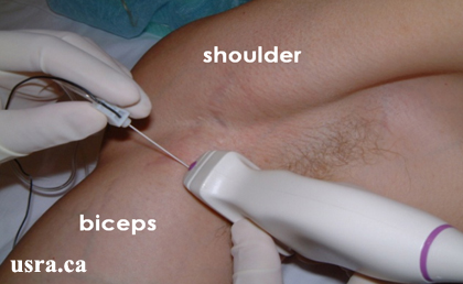
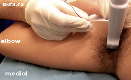
  • Position the patient supine and abduct the arm to 90 degrees.
  • After skin and transducer preparation (see transducer preparation section), place a linear 38-mm, high frequency 10-12 MHz transducer firmly in the transverse plane along the axillary crease to obtain the best possible transverse view of the brachial plexus.
  • Optimize machine imaging capability. Select appropriate depth of field (within 1-2 cm), focus range (usually within 1 cm) and gain.
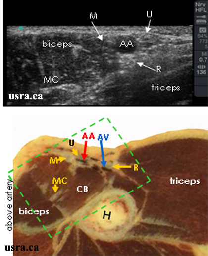
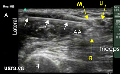
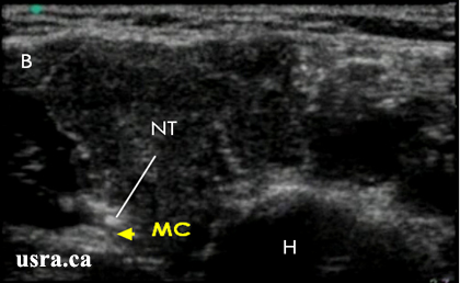
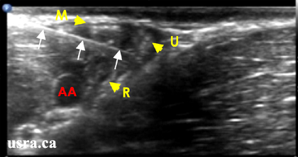
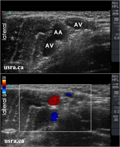
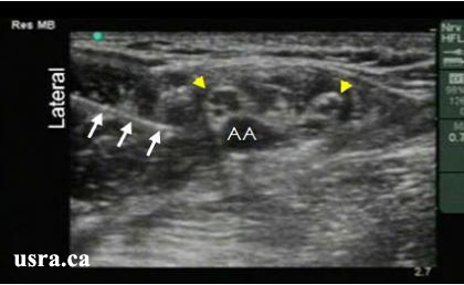
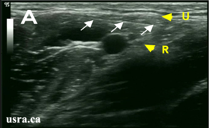
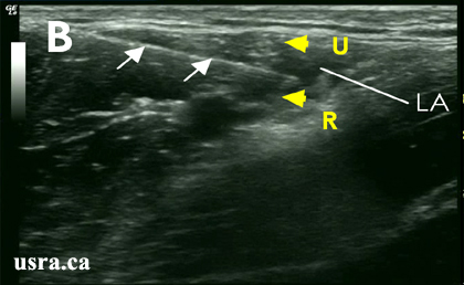
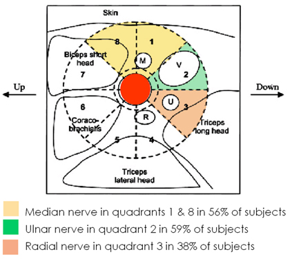
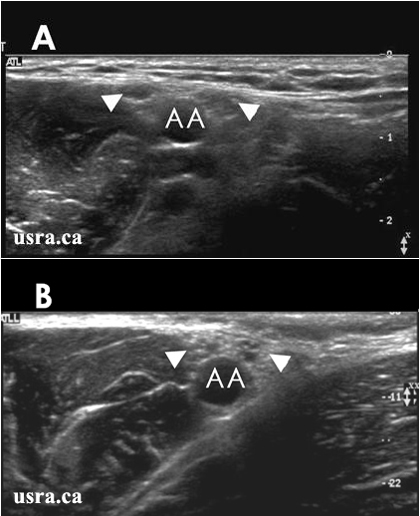
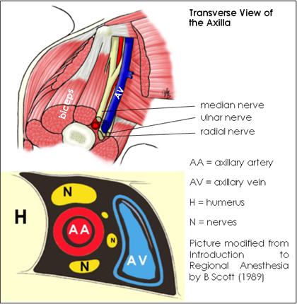
  • Visualize the median, ulnar, radial and musculocutaneous nerves in the transverse view(short axis). Nerves in the axilla have mixed echogenicity and a honey comb appearance (representing a mixture of hypoechoic nerve fasiscles and hyperechoic connective tissues). The nerves are round or oval, and are located next to the axillary artery and vein(s).

Anatomical Correlation

AA and AV = axillary artery and vein Box = scanned area
CB = coracobrachialis muscle H = humerus
M = median nerve MC = musculocutaneous nerve
R = radial nerve U = ulnar nerve

Nerve Localization

  • Perform a systematic anatomical survey of structures from superficial to deep and from the region above the axillary artery to below the artery.
  • Relieve transducer pressure partially on the skin to visualize the compressible axillary vein(s).
  • Identify the triceps, biceps and coracobrachialis muscles surrounding the artery.
  • Identify the humerus deep to the muscles.
  • The median, ulnar and radial nerves are situated around the axillary artery and outside of the muscle layers. They often have a honey comb appearance and are heterogeneous in echogenicity. Expect to find these nerves superficially (often within 1 cm from the skin surface).
  • Move the transducer proximally towards the axilla and distally towards the elbow to appreciate the course of each nerve (nerve tracing).
  • Of these 3 nerves, the radial nerve is often most difficult to locate. It frequently lies deep to the ulnar nerve.
  • Identify the musculocutaneous nerve (hyperechoic), which lies most commonly in the plane between the biceps and coracobrachialis muscles.

Needle Insertion Approach

In Plane Approach

  • Ultrasound guided axillary block is considered a BASIC skill level block because this is a superficial block.
  • Insert a 5 cm 22 G insulated needle parallel to the long axis of the transducer inline with the ultrasound beam as seen in picture below.

  • Observe the transverse view of the terminal branches on the ultrasound monitor. In cross section, nerves appear as round or oval shaped hypoechoic nodules with hyperechoic rims and internal echoes.
  • Also easily identified is the pulsatile axillary artery which is hypoechoic. The axillary veins are often not seen because they are compressed by the ultrasound transducer.
  • The needle should be inserted at a shallow angle because terminal branches of the brachial plexus in the axilla are superficial. As the needle travels in the same plane as the ultrasound beam, the path of advancement can be visualized in real-time as the needle approaches the target nerves.
  • Determine the identity of each individual nerve by electrical stimulation if desired. It is well known that nerves can occupy variable locations around the axillary artery.
  • Block the musculocutaneous nerve separately as it branches in the coracobrachialis muscle.
A. Needle in contact with the median nerve.

Arrows = block needle
AA = axillary artery
H = humerus
M = median nerve
R = radial nerve
U = ulnar nerve
B. The musculocutaneous nerve is blocked separately.

H = humerus
MC = musculocutaneous nerve
NT = needle tip

Blocking The Radial Nerve

  • The radial nerve is usually located deep to the ulnar nerve in the proximal axilla. When the transducer is moved more distally, the radial nerve descends and disappears under the triceps muscle. The radial nerve can be visualized again as it travels posteriorly around the humeral shaft.
  • It is advantageous to target and anesthetize the radial nerve first and then inject around the 2 more superficial nerves (median and ulnar) nerves as the needle is withdrawn.


  • The needle is inserted deep to contact the radial nerve (R) before local anesthetic injection. Notice that the needle is advanced past the superficial median nerve (M) and positioned between the ulnar (U) and radial nerves before injection.

Out of Plane Approach [Link to top]

  • This is a common alternative for needle insertion in the axillary region. Constant injection of a small quantity of fluid (e.g., local anesthetic) is recommended to locate the needle tip at the time of needle advancement. Otherwise the needle tip may have traversed beyond the superficial target nerves without the operator's knowledge.


  • The transducer is positioned in the right axillary region; note the needle is placed perpendicular to the long axis of the transducer.

Finding The Axillary Vein(s)

  • It is important to identify the axillary vein(s) using Color Doppler or Color Power Doppler to avoid unintentional intravascular injection.
  • The axillary veins are often located at the 12 to 2 o'clock and 7 to 9 o'clock locations.
  • Also, the vein often separates the median and ulnar nerves.
  • The vein(s) should be compressed during local anesthetic injection.
  • Detection of an intravascular injection is an important advantage of ultrasound. Intravascular injection is suggested by a lack of local anesthetic spread (a hypoechoic collection) at the time of injection.

Local Anesthetic Injection

  • Inject 10-15 mL of local anesthetic at each nerve location. The minimum effective local anesthetic volume for axillary block has not been determined. Nerve visualization usually becomes increasingly difficult after local anesthetic injection.
Arrows = needle
Arrowheads = nerves
AA = axillary artery
  • It is also important to ensure local anesthetic spread around each individual nerve (median, radial, ulnar and musculocutaneous nerves) at the time of injection. In the opinion of the authors, a perivascular injection around the axillary artery does not guarantee local anesthetic spread around each individual nerve and is not considered adequate to ensure complete nerve blockade. This observation lends support to the concept of septae within the axillary sheath.
  • Improper local anesthetic spread is often indicated by restricted spread to only one side of the nerve (ulnar nerve, U, in figure B below) and is not circumferential. Alternatively, the spread is seen moving away from the nerve suggesting an injection outside the perineural sheath. Circumferential spread is seen in figure C after needle penetration through the fascial sheath.
A. The needle between ulnar (U) and radial (R) nerves

Arrows = block needle
LA = local anesthetic
R = radial nerve
U = ulnar nerve
B. Local anesthetic spread away from ulnar nerve (U)

Arrows = block needle
LA = local anesthetic
R = radial nerve
U = ulnar nerve
C. Local anesthetic spread around ulnar nerve (U)

Arrows = block needle
LA = local anesthetic
R = radial nerve
U = ulnar nerve

Clinical Pearls

Nerve Localization: Anatomical Variations


Ultrasonographic Findings of Variation in Nerve Location Around the Axillary Artery Picture modified from Anesth Analg 2001;92:1271-5.

Median and ulnar nerves (arrowheads) are farther apart in figure A than in B.
AA = axillary artery

Catheter Insertion

Continuous axillary nerve block is generally not recommended due to easy catheter dislodgment. Supraclavicular and infraclavicular catheters are better choices to provide continuous brachial plexus anesthesia and analgesia below the arm.

Image Gallery

Needle and Local Anesthetic Injection Around Individual Nerves

Pre-injection

Arrows = block needle
AA = axillary artery
M = median nerve
U = ulnar nerve
R = radial nerve
Injection # 1
Local anesthetic is first injected deep around the radial nerve (R).

Arrows = block needle
AA = axillary artery
M = median nerve
U = ulnar nerve
Injection # 2
The needle is pulled back to block the superficial median nerve (M).

Arrows = block needle
AA = axillary artery
U = ulnar nerve
Injection # 3
The needle is redirected to block the superficial ulnar nerve (U).

Arrows = block needle
AA = axillary artery
M = median nerve
Post-injection
All 3 nerves are highlighted by local anesthetic after injection. Scan proximally and distally to assess the extent of spread.

M = median nerve
R = radial nerve
U = ulnar nerve

Video Gallery

Selected References

  • Duggan E, Brull R, Lai J, Abbas S: Ultrasound-guided brachial plexus block in a patient with multiple glomangiomatosis. Reg Anesth.Pain Med. 2008; 33: 70-3.
  • Casati A, Danelli G, Baciarello M, Corradi M, Leone S, Di Cianni S, Fanelli G: A prospective, randomized comparison between ultrasound and nerve stimulation guidance for multiple injection axillary brachial plexus block. Anesthesiology 2007; 106: 992-6.
  • Chan VW, Perlas A, McCartney CJ, Brull R, Xu D, Abbas S: Ultrasound guidance improves success rate of axillary brachial plexus block. Can.J.Anaesth. 2007; 54: 176-82.
  • Bigeleisen PE. Nerve puncture and apparent intraneural injection during ultrasound-guided axillary block does not invariably result in neurologic injury. Anesthesiology 2006; 105: 779-83.
  • Sites BD, Beach ML, Spence BC, Wiley CW, Shiffrin J, Hartman GS, Gallagher JD: Ultrasound guidance improves the success rate of a perivascular axillary plexus block. Acta Anaesthesiol.Scand. 2006; 50: 678-84.
  • Perlas A, Niazi A, McCartney C, Chan V, Xu D, Abbas S. The sensitivity of motor response to nerve stimulation and paresthesia for nerve localization as evaluated by ultrasound. Reg Anesth Pain Med 2006; 31: 445-450.
  • Gray A T, Schafhalter-Zoppoth I. "Bayonet artifact" during ultrasound-guided transarterial axillary block. Anesthesiology 2005; 102: 1291-1292.
  • Schwemmer U, Markus C K, Greim C A, Brederlau J, Roewer N. Ultrasound-guided anaesthesia of the axillary brachial plexus: efficacy of multiple injection approach. Ultraschall Med 2005; 26: 114-119.
  • Soeding P E, Sha S, Royse C E, Marks P, Hoy G, Royse A G. A randomized trial of ultrasound-guided brachial plexus anaesthesia in upper limb surgery. Anaesth Intensive Care 2005; 33: 719-725.
  • Bigeleisen P E. The bifid axillary artery. J Clin Anesth 2004; 16: 224-225.
  • Retzl G, Kapral S, Greher M, Mauritz W. Ultrasonographic findings of the axillary part of the brachial plexus. Anesth Analg 2001; 92: 1271-1275.
  • Guzeldemir M E, Ustunsoz B. Ultrasonographic guidance in placing a catheter for continuous axillary brachial plexus block. Anesth Analg 1995; 81: 882-883.
  • Ting P L, Sivagnanaratnam V. Ultrasonographic study of the spread of local anaesthetic during axillary brachial plexus block. Br J Anaesth 1989; 63: 326-329.

Share to Facebook Share to Twitter More...