Youtube

Advancing the Science of Ultrasound Guided Regional Anesthesia and Pain Medicine

Intercostal Nerve Block

Introduction

Intercostal nerve block with local anesthetic and/or steroid is performed for the relief of chest wall pain secondary to surgery, trauma, cancer, and post herpetic neuralgia. Chemical neurolysis and cryoablation of the intercostal nerves has been performed for treatment of persistent post thoracotomy pain.

Anatomy

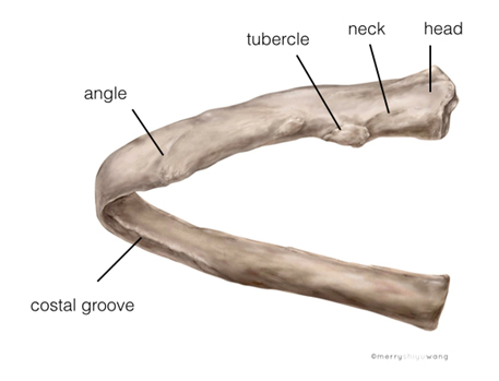
The intercostal space (ICS) is the space between 2 adjacent ribs of the thoracic cage. A typical rib (rib 3rd to 10th) has the following structures: head, neck, tubercle, angle and body Figure 1). Typically a rib courses oblique as it curves laterally and anteriorly and so is the intercostal space. Note that the costal groove in the inferior border of the rib ends as it courses more anteriorly (Figure 1).

Figure 1. Anatomy of a rib


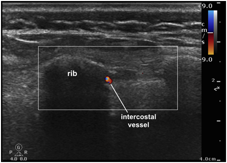
There are 11 ICSs on each side and the content of each ICS includes: intercostal muscles, intercostal membranes, intercostal nerves (ICNs) and intercostal vessels (Figure 2). Deep to the intercostal space is the parietal pleura. Lateral to the costal angle, the ribs and ICSs are superficial thus easily palpable on the back.

Figure 2. Posterior view of the intercostal nerves running through the posterior intercostal spaces between two ribs. The erector spinae muscle is completely removed and the external intercostal muscles are partially removed in this dissection


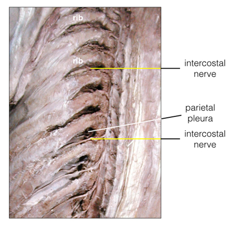
There are 11 pairs of ICNs (T1-T11) which are anterior divisions of the thoracic spinal nerves. They course through the intercostal spaces accompanied by the intercostal vessels. They provide sensory and motor innervation to the thoracic and abdominal wall and sensory innervation to the parietal pleura and peritoneum.

At the posterior axillary line, the ICNs divide in main and collateral branches. The former is found near the inferior border of the upper rib of an ICS and the latter follows the superior border of the lower rib (note that the intercostal vessels also branch in the same pattern.

Moreover, approximately at the midaxillary line, the ICNs give off the lateral cutaneous branches which further divide into anterior and posterior cutaneous terminal branches. To ensure complete sensory blockade, it is therefore important to anesthetize the intercostal nerves at or posterior to the posterior axillary line.

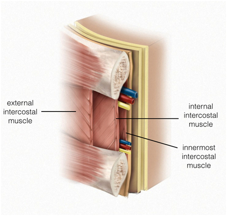
Lateral to the costal angle, there are 3 layers of muscles? external intercostal muscle (EICM), the internal intercostal muscle (IICM) and the innermost intercostal muscle (IMICM) (Figure 3). The innermost intercostal muscle, also called the intercostalis intimus muscle, is a flimsy innermost muscle layer which readily permits local anesthetic diffusion. Deep to the innermost intercostal muscle is the parietal pleura. From the costal angle onwards, the main ICNs and vessels, grouped as a neurovascular bundle, are arranged in the vein, artery and nerve (VAN) orientation from superior to inferior. Note that this constant arrangement does not apply for the collateral bundle. Both neurovascular bundles are found between the internal intercostal muscle and innermost intercostal muscle but the main bundle is found within the costal groove or near the inferior margin of the upper rib of an ICS while the collateral bundle is found at the superior border of the lower rib.

Figure 3. Schematic diagram of the content of the anterior ICS (anterior to the posterior axillary line) after the IC neurovascular bundle has divided into the main and collateral branches


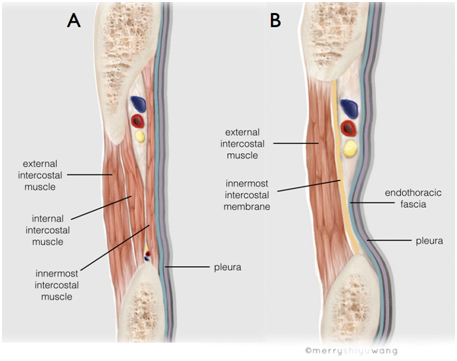
Medial to the costal angle, there is only 1 layer of muscle, the external intercostal muscle and an internal intercostal membrane (IICMe, Figure 4). There is no internal intercostal muscle. The intercostal neurovascular bundle runs between the ribs, deep to the internal intercostal membrane and superficial to the endothoracic fascia (ETF). There is no clear intercostal groove in this location. Deep to the endothoracic fascia is the parietal pleura.

Figure 4. Content of the posterior ICS (posterior to posterior axillary line) lateral to the costal angle (A) and medial to the costal angle (B). It shows the location of the intercostal nerve and vessels in relationship to the intercostal musculature and pleura


Sonoanatomy

In the majority of cases, it is not possible to visualize sonographically the ICNs in the ICSs lateral to costal angles. In this region, the ICNs are very close to the rib thus visibility is impeded by bone shadow.

Also, the nerves are progressively smaller when course beyond the costal angles (see dissection in Figure 4 and sonograms in Figures 5 and 6). The intercostal muscles, ribs and pleura are important landmarks that are easily recognized leading to the intercostal nerves (see ultrasound images).

Figure 5. Sonogram showing the intercostal space lateral to the costal angle


* denotes fascial layer between the external and internal intercostal muscles; the intercostal nerve is not visualized in this region

Figure 6. Sonogram showing the intercostal space medial to the costal angle


* location of intercostal nerve (not visualized)

Scanning Technique

Place the patient in the prone or sitting position. Alternatively, place the patient in the lateral decubitus position with the side to be blocked uppermost.

Scan the posterior intercostal space laterally (lateral to the costal angle) but posterior to the posterior axillary line where the neurovascular bundle has not yet divided (see Anatomy section above).

After skin and transducer preparation, place a 38 mm linear transducer with the appropriate frequency range (10-12 MHz) oblique in the back at right angle to two palpable ribs (Figure 7). Note that the obliquity of transducer position changes according to the level of block.

Figure 7. Transducer positioned oblique and lateral to the costal angle


Optimize machine imaging capability; select appropriate depth of field, focus range and gain.

Start scanning at the inferior angle of the scapula which corresponds to approximately the 7th ICS when the hand is hanging down on the side. Note that when scanning the 2nd to 7th ICSs, the scapula has to be moved laterally (by hanging the arm on the side or having the arm reached over to the contralateral shoulder) to allow scanning lateral to costal angles.

Nerve Localization

Perform a systematic anatomical survey from superficial to deep in the posterior intercostal space lateral to the costal angle.

It is often not possible to visualize the intercostal nerves in the intercostal spaces (see explanation above). However, the vessels within the collateral neurovascular bundle located in the upper margin of the rib below may be visible using Color (Power) Doppler.

The intercostal neurovascular bundle (both main and collateral) is expected to lie between the internal intercostal muscle and the innermost intercostal muscle. The innermost intercostal muscle is a flimsy muscle layer that is not always clearly visualized under ultrasound. For this reason, the internal intercostal muscle is the important sonographic muscle layer to identify that will lead to the intercostal neurovascular bundle.

In contrast, the parietal pleura deep to the innermost intercostal muscle is readily recognized as a very bright, hyperechoic line that slides with respiration. There are also associated air artifacts (comet tail sign) deep to the reflex of the parietal pleura.

With the lateral approach, the indirect image target for the intercostal nerve is the internal intercostal muscle since we know that the ICN is between the internal intercostal muscle and the innermost intercostal muscle.

Color Doppler may visualize the intercostal vessels in the inferior intercostal groove (Figure 8).

Figure 8. Color Doppler lateral to the costal angle showing the intercostal vessels


Needle Insertion Approach
Ultrasound guided posterior intercostal nerve block is considered a BASIC skill level block (Level 1). The only major risk is pneumothorax should the needle puncture the pleura.

For posterior intercostal nerve block, the ideal needle insertion site is the ICS lateral to the costal angle and posterior to the posterior axillary line (because the neurovascular bundle has not yet divided; see Anatomy).

An out-of-plane approach, with direct access to the intercostal space, as well as an in?plane technique is possible. With the in-plane approach, a transducer with a small foot print is required (Figure 9). The in-plane approach has the advantage of visualizing the block needle more clearly but the passage may be partially obstructed by the rib one space below. This is true especially in case of narrow ICSs.

Figure 9. In plane needle advancement from caudad to cephalad


With either needle approach, it is important to hydrodissect repeatedly during needle advancement in the tissue plane before reaching the internal intercostal muscle. This will facilitate visualization of the needle tip and identify the correct tissue layer.

With the patient lying prone, a 22 G, 3.5 or 5 cm needle is advanced to penetrate the external and internal intercostal muscles (Figure 10). The optimal target needle endpoint is a location just within the internal intercostal muscle to assure that the needle tip remains superficial to the parietal pleura. Again, the innermost intercostal muscle is not always visualized thus is not a useful landmark to guide injection. The clearly visible internal intercostal muscle serves an indirect sonographic target for needle insertion.

Figure 10. Sonogram showing needle using an in plane insertion approach


arrows = block needle

The needle should not traverse the parietal pleura to avoid pneumothorax.

Perform a post block ultrasound scan to rule out pneumothorax. The absence of comet tail artifacts and sliding pleura indicates pneumothorax.

Local Anesthetic Injection

The first goal is to block ICNs lateral to the costal angle and posterior to the posterior axillary line.

The second goal is to inject local anesthetic deep to the internal intercostal muscle and visualize depression of the parietal pleura during injection. Sonographically, one aims to inject within the internal intercostal muscle (explanation in the Anatomy section).

Injection of 2 mL of local anesthetic is usually sufficient for intercostal nerve block.

Hariharan Shankar, MD
Associate Professor Department of Anesthesiology
Medical College of Wisconsin Milwaukee WI, USA


Video showing needle advancement, hydrodissection and depression of the pleura by local anesthetic injection

Clinical Pearls

Anatomy

1. Intercostal Muscle Orientation


The external intercostal muscle (EICM) fibers run in an oblique plane from supero-posterior to infero-anterior while the internal intercostal muscle (IICM) fibers run in the opposite direction, from infero-posterior to supero-anterior. Thus the external and internal muscle fibers run at right angles to each other in the intercostal space.

To capture a discrete view of the EICM and IICM within an ICS, it is best to orient the transducer and the ultrasound beam inline with the orientation of the respective muscle fiber (see images below). The EICMs and IICMs are only visualized as 2 separate muscle layers when the transducer is placed lateral to the costal angle. Again note that there is no IICMs medial to costal angle.

For the ICN block procedure, however, the transducer is best positioned at right angle to the rib and not in the same orientation of the intercostal muscle fiber.

Avoid improper scanning technique. That is, do not position the ultrasound transducer in a perfectly sagittal or transverse orientation.

2. Scapula

The inferior angle of the scapula is an important external landmark for counting the level of the posterior intercostal space prior to ICN block. Generally speaking, this corresponds to the 7th intercostal space when the hand is hanging down on the side. When the hand is placed over the head or reached over the contralateral shoulder, this corresponds approximately to the 6th intercostal space.

The intercostal spaces are more difficult to access from the 2nd to the 7th rib because of the overlying scapula. Again, placing the hand over the head, contralateral shoulder or bringing both shoulders forward, helps to move the scapula more laterally.

Needling

1. Needling Medial to the Costal Angle (an Alternative Approach)


Medially, the ribs and the posterior intercostal spaces are more difficult to access because they are deep to the overlying paraspinal muscles, i.e. mainly the erector spinae, rhomboids and trapezius muscles as well as the latissimus dorsi muscles inferiorly. Again medial to the costal angle, there is only 1 layer of muscle, the external intercostal muscle and an internal intercostal membrane. There is no internal intercostal muscle.

The intercostal neurovascular bundle runs deep to the internal intercostal membrane and superficial to the endothoracic fascia. Deep to the endothoracic fascia is the parietal pleura. In essence, the ICN medial to the coastal angle is literally in direct contact with the pleura. Sonographically, the endothoracic fascia is usually not visualized.

An advantage of performing a medial needling approach is the possibility of directly visualizing the ICN (see US image in Figure 5). However, because of the needle tip proximity to the pleura and therefore a narrow margin of safety, the medial needle insertion technique is reserved for the advanced experienced practitioners.

Furthermore, nerve visualization medial to the costal angle may be impaired or even impossible in obese or athletic individuals. A lower frequency transducer may be required.

2. Difficulty Needle Visualization

As visualization of a 25 G needle may be difficult with this technique a larger needle may be chosen. One alternative is to use frequent hydrodissection as the needle is advanced.

The use of echogenic needles may permit better visualization.

While performing hydrodissection, it is important to remember that a large volume injection may obscure the muscular landmarks. A small volume injection with 0.1- 0.2 mL is recommended for needle tip localization.

3. Post Procedure Scanning

It is always good practice to perform a post procedure scan to ensure the integrity of pleura by observing the sliding sign and the comet tail sign.

Local Anesthetic Injection

1. Incomplete Block


Misinterpretation of the muscle layers, external intercostal muscle vs. the internal intercostal muscle, will lead to incorrect positioning of the needle tip and block failure. Hydrodissection is recommended to distinguish the intercostal muscle layers.

Inappropriate block location is another reason for incomplete block. To provide complete chest wall anesthesia/analgesia, it is important to perform the block at or posterior to the posterior axillary line before lateral cutaneous branching of the intercostal nerve.

As there is likely to be some cross coverage, blocking the adjacent levels ensures complete analgesia.

It is important to remember that injection as close as possible to the target is critical for increased efficacy.

Selected References

  • Patel SI, Joshi MY. Neurostimulation with ultrasound guidance for intercostal nerve block. PM R 2013;5:903-5.
  • Vandepitte C, Gautier P, Bellen P, Murata H, Salviz EA, Hadzic A. Use of ultrasound-guided intercostal nerve block as a sole anaesthetic technique in a high-risk patient with Duchenne muscular dystrophy. Acta Anaesthesiol Belg 2013;64:91-4.
  • Narouze SN, Provenzano D, Peng P, Eichenberger U, Lee SC, Nicholls B, Moriggl B. The American Society of Regional Anesthesia and Pain Medicine, the European Society of Regional Anaesthesia and Pain Therapy, and the Asian Australasian Federation of Pain Societies Joint Committee Recommendations for Education and Training in Ultrasound-Guided Interventional Pain Procedures. Reg Anesth Pain Med 2012;37:657-64.
  • Stone MB, Carnell J, Fischer JWJ, Herring AA, Nagdev A. Ultrasound-guided intercostal nerve block for traumatic pneumothorax requiring tube thoracostomy. Am J Emerg Med 2011;29:697.e1-2.
  • Peng PW, Narouze S. Ultrasound-guided interventional procedures in pain medicine: a review of anatomy, sonoanatomy, and procedures: part I: nonaxial structures. Reg Anesth Pain Med 2009;34:458-474.
  • Gofeld M. Ultrasonography in pain medicine: a critical review. Pain Pract 2008;8:226-24027.

Share to Facebook Share to Twitter More...