Youtube

Advancing the Science of Ultrasound Guided Regional Anesthesia and Pain Medicine

Cervical Sympathetic Trunk Block

Introduction

Stellate ganglion block is often the term used to describe blockade of the cervical sympathetic trunk. This block is performed for the following indications and purposes for diagnosis or treatment:

  1. provide sympathetic vasodilatation to relieve vasospasm and increase vascular flow; it is indicated for treatment of acute post traumatic or post microvascular surgery vasospasm, frostbite in the upper extremity, Raynaud's syndrome, chronic obliterative arterial diseases,
  2. relieve sympathetically mediated neuropathic pain in the upper extremity such as complex regional pain syndrome types I and II
  3. relieve sympathetically mediated visceral pain e.g., refractory angina pectoralis
  4. others e.g., hyperhidrosis

Anatomy

The cervical sympathetic trunk, one on each side of the vertebral bodies, carries sympathetic fibers to the head and neck structures. The cervical sympathetic trunk has 3 ganglia: superior, middle and inferior ganglia (Figure 1).

Figure 1. Coronal section of the neck showing the cervical sympathetic trunk and its relation to the prevertebral muscles on each side


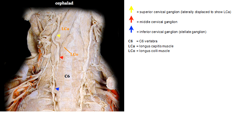
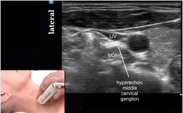
The superior cervical ganglion containing C1-4 fibers is usually found opposite the C2 and C3 vertebrae. The middle cervical ganglion containing C5 and C6 fibers is often found opposite the C6 vertebrae. It is the smallest of the 3 cervical ganglia and may be absent. The inferior cervical ganglion containing C7 and C8 fibers is often located between the base of the C7 transverse process and the neck of the 1st rib, on the medial side of the costocervical artery.

Movie 1. Cervical sympathetic ganglia and trunk anatomy


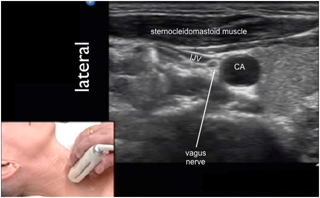
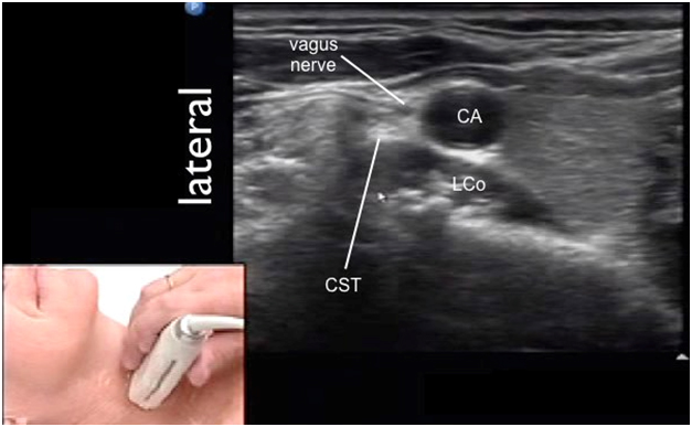
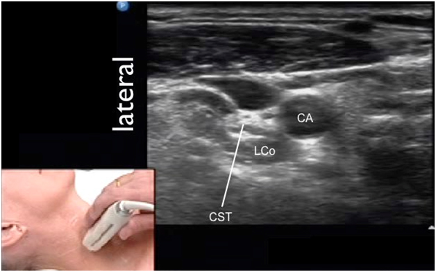
When the inferior cervical ganglion is fused with the T1 ganglion, this cervicothoracic ganglion is called the stellate ganglion. It is often situated posterior to the vertebral artery and the subclavian artery and lateral to the trachea, esophagus and the longus colli muscle. The post-ganglionic fibers of the stellate ganglion provide sympathetic innervation to the upper limbs.

Movie 2. Stellate ganglion anatomy


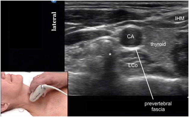
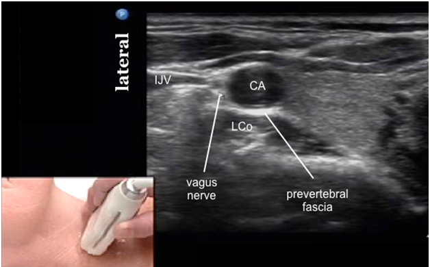
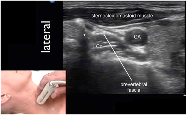
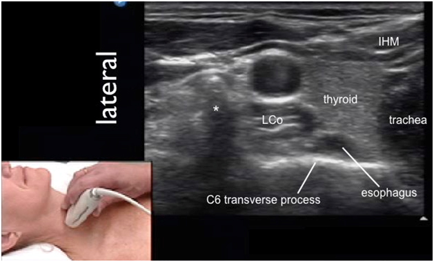
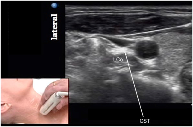
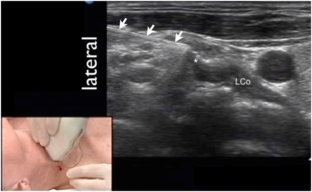
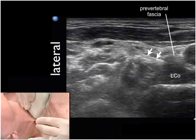
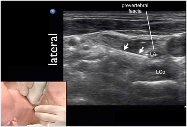
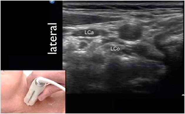
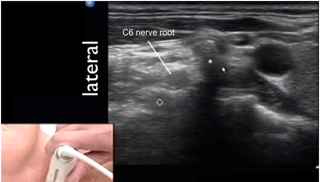
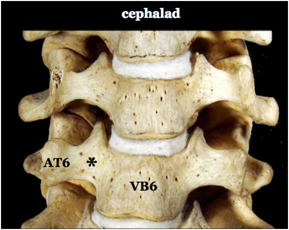
The prevertebral fascia is a layer of deep cervical fascia that envelops the prevertebral muscles (longus colli and longus capitis muscles) that attach to the cervical vertebral bodies and transverse processes (Figure 2). The cervical sympathetic trunk runs through the prevertebral fascia thus this important structure influences the extent of local anesthetic spread during stellate ganglion block. Success of the cervical sympathetic block relies on proper local anesthetic deposit deep to the prevertebral fascia.

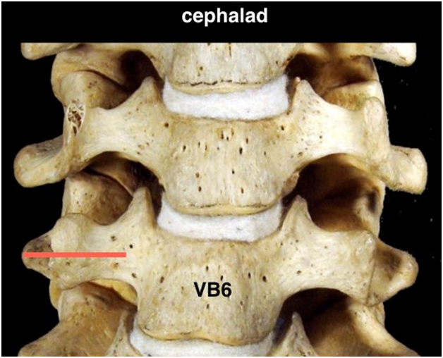
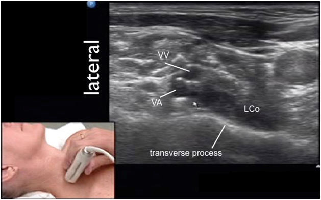
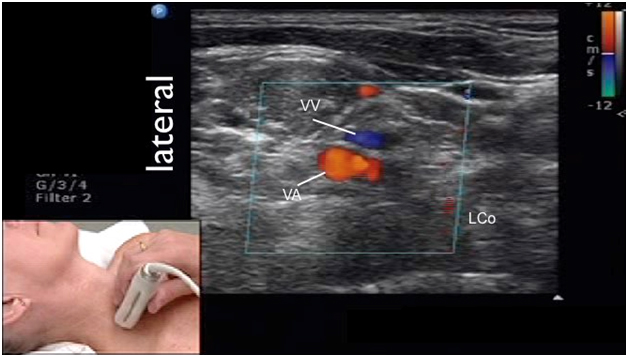
Figure 2. Anterior view of the cervical spine showing the C6 transverse process

AT6 = anterior tubercle of C6 transverse process
VB6 = C6 vertebral body
* = groove between TP and VB6 where longus colli muscle is located

Movie 3. C6 transverse process anatomy

Sonoanatomy

In the transverse view, although the cervical sympathetic trunk is expected to lie between the longus colli muscle and the prevertebral fascia, it may not be easily visualized as a distinct structure until local anesthetic is injected to separate the longus colli muscle from the prevertebral fascia (Figure 3).

Figure 3. Sonogram at the C6 vertebra level showing the expected location of the cervical sympathetic trunk

* = anterior tubercle of C6 transverse process
CA = carotid artery
IHM = infra hyoid musculature
LCo = longus colli muscle

The cervical sympathetic trunk may appear hyperechoic. This is likely the middle cervical ganglion at C6 location (Figure 4).

Figure 4. Sonoanatomy of the cervical sympathetic trunk

IJV = internal jugular vein
LCo = longus colli muscle

The neurovascular bundle adjacent to the cervical sympathetic trunk comprises the carotid artery (CA), internal jugular vein (IJV) and the vagus nerve as shown in Figure 5.

Figure 5. Sonoanatomy showing the vagus nerve near to the cervical sympathetic trunk

CA = carotid artery
IJV = internal jugular vein
LCo = longus colli muscle

Scanning Technique

Position the patient lateral decubitus (with the block side up) or supine.

After skin and transducer preparation, place a linear 38-mm high frequency 10-12 MHz transducer on the skin surface to obtain a best possible transverse view of the neck (Figure 6).

Figure 6. A linear transducer positioned over neck


Optimize machine imaging capability by selecting the appropriate depth of field (usually within 2-4 cm), focus range and gain.

The transducer should be at the level of the C6 transverse process (Figure 7).

Figure 7. Optimal transducer position (red line) shown on a skeleton

Nerve Localization

Perform a systematic anatomical survey.

External palpable landmarks surrounding the cervical sympathetic trunk that are helpful reference points when initiate scanning are:

  1. sternocleidomastoid muscle;
  2. cricoid cartilage (C6 level);
  3. carotid artery;
  4. the carotid tubercle or anterior tubercle of the C6 transverse process and
  5. trachea (midline).

Helpful internal sonographic landmarks to be recognized at the C6 level are:

  1. sternocleidomastoid muscle;
  2. cricoid cartilage (C6 level);
  3. carotid artery;
  4. anterior tubercle of the C6 transverse process);
  5. infra-hyoid musculature;
  6. longus colli muscle;
  7. trachea;
  8. thyroid gland;
  9. vertebral body; and
  10. esophagus.

First identify the superficial triangular shaped sternoclei-domastoid muscle (SCM).

Then identify the neurovascular landmarks within the carotid sheath which are the carotid artery (CA), internal jugular vein and the vagus nerve. They are deep to the sternocleidomastoid muscle (Figure 8).

Figure 8. Sonoanatomy of neurovascular landmarks within the carotid sheath

CA = carotid artery
IJV = internal jugular vein

Deep to the carotid artery is the longus colli muscle (LC) and the overlying prevertebral fascia (Figure 9). The longus colli muscle lies anterior to the C6 vertebral body. This is the reference muscle for cervical sympathetic block at the C6 level.

Figure 9. Sonoanatomy of longus colli muscle and prevertebral fascia

* = anterior tubercle of C6 transverse process
CA = carotid artery
LCo = longus colli muscle

Medial to the carotid artery is the thyroid gland (Th), a homogeneously hyperechoic organ. Deep to the thyroid gland is the esophagus (E) which is more readily visible especially on the left side of the neck (Figure 10). The deepest structure is the transverse process (TP, a hyperechoic line with a bony acoustic shadow beneath. Medial to the thyroid gland is the cricoid cartilage and the trachea (T). Both are easily palpable by hand and visible under ultrasound.

Figure 10. Sonoanatomy of internal sonographic landmarks at C6 level

* = anterior tubercle of C6 transverse process
IHM = infra hyoid musculature
LCo = longus colli muscle

Lateral to the carotid artery at the level of C6 is the C6 transverse process with the anterior tubercle (AT) which is more prominent than the posterior tubercle. Both tubercles are seen as bony acoustic shadows. Simply follow the hyperechoic line of the vertebral body laterally to the prominent anterior tubercle of the C6 transverse process.

The hyperechoic cervical sympathetic trunk is expected to lie anterior to the longus colli muscle, posterior to the carotid artery and medial to the anterior tubercle of the C6 transverse process (Figure 11). This is likely where the middle cervical ganglion or trunk is situated. This is not the stellate ganglion which is located more inferiorly (between the C7 transverse process and the first rib).

Figure 11. Sonoanatomy of cervical sympathetic trunk

CST = cervical sympathetic trunk, most likely the middle cervical ganglion, NOT the stellate ganglion which is at C7/T1 level
LCo = longus colli muscle

The prevertebral fascia overlying the longus colli muscle is thin and may appear as a hyperechoic line. However it is not easily visualized under ultrasound most of the time. The prevertebral fascia becomes visualized once it is separated from the longus colli muscle during injection of local anesthetic.

Now identify the optimal site for CST block at the C6 level.

Needle Insertion Approach

Ultrasound guided cervical sympathetic trunk block is considered a BASIC skill level block.

The goal is to block the cervical sympathetic trunk at approximately the C6 level which corresponds to approximately the middle cervical ganglia level. Local anesthetic deposited at C6 will spread caudad to block the stellate ganglion.

To approach the cervical sympathetic trunk at the C6 level, it is advisable to turn the head to the contralateral side of the block. This will bring the cervical sympathetic trunk and the longus colli muscle lateral to the carotid artery, and the vagus nerve.

We recommend an in-plane lateral to medial needle approach (Figure 12).

Figure 12. Transducer and patient position for ultrasound guided cervical sympathetic block


The needle approaching from lateral to medial will first contact the anterior tubercle of the C6 transverse process (Figure 13) and then enter the prevertebral fascia (Figure 14).

Figure 13. Needle advancement to contact C6 anterior tubercle

arrows = needle
* = anterior tubercle of C6 transverse process
LCo = longus colli muscle

Figure 14. Needle entering the prevertebral fascia

arrows = needle
LCo = longus colli muscle

Local Anesthetic Injection

The goal is to inject 5 mL of local anesthetic deep to the prevertebral fascia (or called the deep cervical fascia) and above the longus colli muscle (Figure 15). Also aim not to inject local anesthetic into the substance of the longus colli muscle.

Figure 15. Local anesthetic deposited deep to the prevertebral fascia

arrows = needle
LA = local anesthetic
LCo = longus colli muscle

Movie 4 Local anesthetic injection


Movie 5 Local anesthetic injection

Clinical Pearls

Longus Capitis Muscle

When scan cephalad to the C6 level, the longus capitis muscle (LCa) becomes visible lateral to the C6 transverse process (Figure 16).

Figure 16. Sonoanatomy of longus capitis muscle when scanned cephalad to C6

LCa = longus capitis muscle
LCo = longus colli muscle

Vertebral Artery and Vein

When scan caudad to the C6 level, the vertebral artery (VA) and vein (VV) are now in view (Figure 17 13). The stellate ganglion is in the vertebro-scalene triangle at this level. The stellate ganglion is posterior to the vertebral vessels thus difficult to access.

Sonogram at the C7 level showing the longus colli muscle (LC), the vertebral artery and vein (VA & VV) and the vertebral body (VB) as shown in Figures 17 (without Color Doppler) and 18 (with Color Doppler). Note that the C6 transverse process is absent in this view.?

Figure 17. Sonogram caudad to C6 showing vertebral artery and vein

LCo = longus colli muscle
VA = vertebral artery
VV = vertebral vein

Figure 18. Sonogram caudad to C6 showing vertebral artery and vein (with Color Doppler)

LCo = longus colli muscle
VA = vertebral artery
VV = vertebral vein

The C6 Nerve Root

The cervical sympathetic trunk is medial to the C6 transverse process while the C6 nerve root is lateral to the anterior tubercle (AT) of C6 (Figure 19). In this picture, the C6 nerve root is seen exiting the neural foramen at the C6 transverse process.

Figure 19. Sonogram showing the C6 nerve root

* = anterior tubercle of C6 transverse process
= posterior tubercle of C6 transverse process

The Effect of Head Turning

The cervical sympathetic trunk and the longus colli muscle are usually located deep to the carotid artery when the head is in neutral position (Figure 20). To access the cervical sympathetic trunk for an ultrasound guided procedure, turn the patient's head to the contralateral side so that the sympathetic trunk and the longus colli muscle are displaced more laterally (Figure 21).

Figure 20. Sonogram showing the cervical sympathetic trunk and longus colli muscle deep to the carotid artery when the head is in neutral position

CA = carotid artery
CST = cervical sympathetic trunk
LCo = longus colli muscle

Figure 21. Sonogram showing the cervical sympathetic trunk and longus colli muscle lateral to the carotid artery when the head is turned to the contralateral position

CA = carotid artery
CST = cervical sympathetic trunk
LCo = longus colli muscle

Vertebro-scalene Triangle

At the vertebro-scalene triangle at C7 or below, the cervicothoracic (stellate) ganglion situated between the C7 transverse process and the first rib is immediately next to the vertebral vessels thus needle access to this region is technically challenging (Figure 22).

Figure 22. Sonogram showing the vertebro-scalene triangle C7 and below

VA = vertebral artery
VV = vertebral vein

Video Gallery

Selected References

  1. Bhatia A, Flamer D, Peng PWH. Evaluation of sonoanatomy relevant to performing stellate ganglion blocks using anterior and lateral simulated approaches: an observational study. Can J Anaesth 2012;59:1040-7.
  2. Gofeld M, Bhatia A, Abbas S, Ganapathy S, Johnson M. Development and validation of a new technique for ultrasound-guided stellate ganglion block. Reg Anesth Pain Med 2009;34:475-9.
  3. Kapral S, Krafft P, Gosch M. Ultrasound imaging for stellate ganglion block: direct visualization of puncture site and local anesthetic spread: a pilot study. Reg Anesth 1995;20:323-8.
  4. Narouze S. Ultrasound-guided stellate ganglion block: safety and efficacy. Curr Pain Headache Rep 2014;18:424.
  5. Nix CM, Harmon DC. Avoiding intravascular injection during ultrasound-guided stellate ganglion block. Anaesthesia 2011;66:134-5.
  6. Ojeda A, Sala-Blanch X, Moreno LA, Busquets C. Ultrasound-guided stellate ganglion block: what about the phrenic nerve? Reg Anesth Pain Med 2013;38:170.
  7. Rastogi S, Tripathi S. Cardiac arrest following stellate ganglion block performed under ultrasound guidance. Anaesthesia 2010;65:1042.
  8. Shankar H, Simhan S. Transient neuronal injury followed by intravascular injection during an ultrasound guided stellate ganglion block. Anesth Pain Med 2013;2:134-7.
  9. Soneji N, Peng PWH. Ultrasound-guided pain interventions - a review of techniques for peripheral nerves. Korean J Pain 2013;26:111-24.
  10. Usui Y, Kobayashi T, Kakinuma H, Watanabe K, Kitajima T, Matsuno K. An anatomical basis for blocking of the deep cervical plexus and cervical sympathetic tract using an ultrasound-guided technique. Anesth Analg 2010;110:964-8.
  11. Yoo SD, Jung SS, Kim H-S, Yun DH, Kim DH, Chon J, Hong DW. Efficacy of ultrasonography guided stellate ganglion blockade in the stroke patients with complex regional pain syndrome. Ann Rehabil Med 2012;36:633-9.
  12. Wei K, Feldmann RE, Brascher A-K, Benrath J. Ultrasound-guided stellate ganglion blocks combined with pharmacological and occupational therapy in Complex Regional Pain Syndrome (CRPS): a pilot case series ad interim. Pain Med 2014;15:2120-7.

Share to Facebook Share to Twitter More...