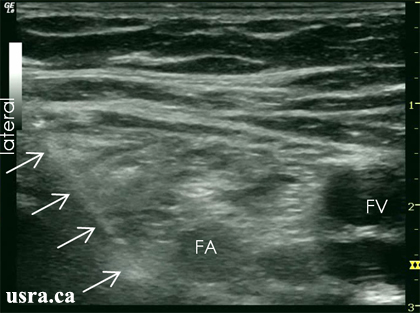
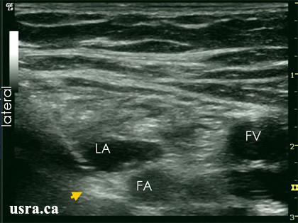
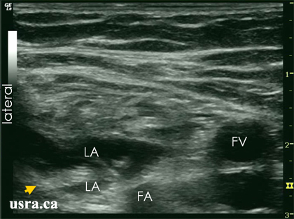
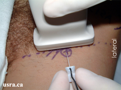
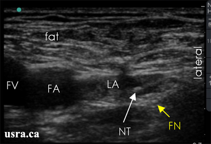
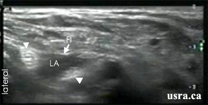
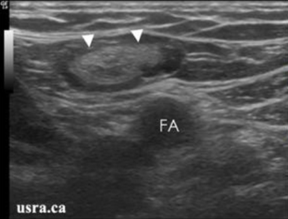
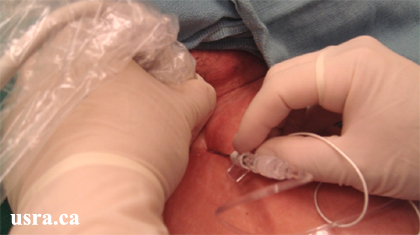
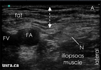
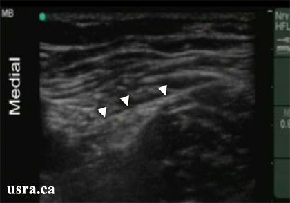
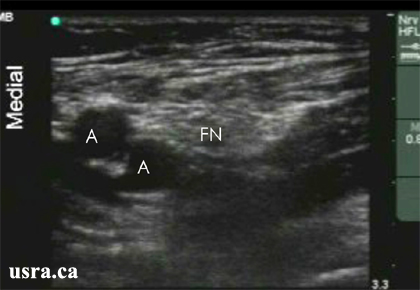
Youtube

Advancing the Science of Ultrasound Guided Regional Anesthesia and Pain Medicine

Femoral Nerve Block

Anatomy

The femoral nerve is one of the major branches of the lumbar plexus. The femoral nerve is consistently lateral to the femoral artery, deep to the fascia iliaca and superficial to the iliopsoas muscle. The anterior approach to block the femoral nerve at the groin (inguinal region) is most commonly performed for knee surgery.

FA = femoral artery
FN = femoral nerve
FV = femoral vein

Picture taken from Colour Atlas of Anatomy: A photographic study of the human body (3rd edition)
FI = fascia iliaca
FL = fascia lata
FS = femoral sheath
IPM = iliopsoas muscle
PECT = pectineus muscle

Scanning Technique

  • Position the patient supine with the leg in the neutral position.
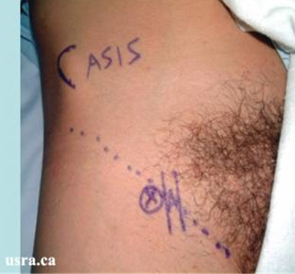
  • Expose the groin and mark the inguinal crease.
  • After skin and transducer preparation, place a transducer with the appropriate frequency range (10-12 MHz) along the inguinal crease. If the femoral artery and nerve are deep (> 4 cm, use a 7 MHZ transducer).
  • Optimize machine imaging capability; select appropriate depth of field (usually within 1-3 cm), focus range and gain.

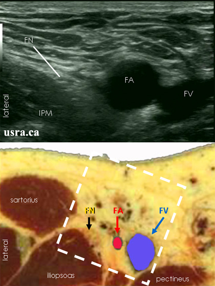
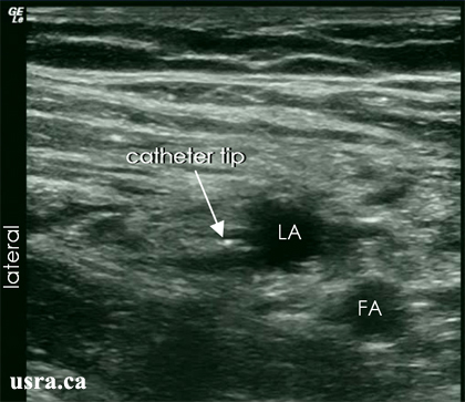
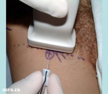
Transducer over right inguinal crease

Anatomical Correlation

Transverse Scan of the Inguinal Region

Box = scanned area
FA = femoral artery
FN = femoral nerve
FV = femoral vein
IPM = iliopsoas muscle

Nerve Localization

  • Perform a systematic anatomical survey from medial to lateral and superficial to deep.
  • The femoral nerve is generally easy to locate in this region.
  • First, identify the femoral artery. If the image shows more than 1 artery, scan more proximally (cephalad) to visualize the artery before the profunda femoris artery branches off.
  • The femoral vein is medial to the artery. The vein may not be visible until the transducer pressure on the skin is lessened.
  • Deep to the femoral vessels is the iliopsoas muscle bulk.
  • The femoral nerve is often found within a triangular hyperechoic region, lateral to the femoral artery and superficial to the iliopsoas muscle.
  • The femoral nerve may be quite thin and flat in this region as the nerve fans out into multiple branches.
  • Note the fascia iliaca (a hyperechoic line) superficial to the femoral nerve and its branches.
  • Inguinal lymph nodes also appear hyperechoic and may be confused with the nerve in the short axis view. To distinguish the two, scan proximally and distally in this region. A nerve is a continuous structure that can be traced while a lymph node is not and can be seen only in a discrete location.

Needle Insertion Approach

In Plane Approach

  • The in plane approach is also commonly used for femoral nerve block by aligning the block needle with the ultrasound beam.
  • With this approach, the needle shaft and tip can be visualized distinctly but it may take a longer time to align the needle with the beam compared to the out of plane approach.
Insertion of a block needle over the left inguinal region using the in plane approach
In plane needle approach showing needle in contact with the femoral nerve

Arrows = block needle
FA = femoral artery
FV = femoral vein
In plane needle approach showing needle in contact with the femoral nerve

Arrowhead = femoral nerve
FA = femoral artery
FV = femoral vein
LA = local anesthetic
In plane needle approach showing needle in contact with the femoral nerve

Arrows = block needle
FA = femoral artery
FV = femoral vein
LA = local anesthetic

Out of Plane Approach

  • Ultrasound guided femoral nerve block is considered a BASIC skill level block because this is a superficial block.
Insert a 5 cm 22 G insulated needle perpendicular to the transducer and the ultrasound beam. In this case, only the cross section of the needle shaft (a white dot) may be observed during needle advancement.
  • It can be technically challenging to track the location of the needle tip during needle insertion without an echogenic tip design. Move the needle tip slightly from side to side or in and out to bring the tip into view.
  • Injection of a small amount of fluid e.g., D5W will expand the femoral triangle and the hypoechoic fluid collection can bring the hyperechoic nerve and the fascia iliaca into view.
  • Identify the femoral nerve branches by electrical stimulation. Aim to evoke patellar contraction.
  • Injection of D5W (1-5 mL) will also intensify the motor response to nerve stimulation.
  • The posterior division of the femoral nerve which innervates the quadriceps muscles is most commonly located on the lateral aspect of the femoral triangle. It is therefore recommended to first point the needle towards the lateral aspect of the femoral triangle under ultrasound guidance.
FA = femoral artery
FN = femoral nerve
FV = femoral vein
LA = local anesthetic
NT = needle tip

Local Anesthetic Injection

  • Once satisfied with needle placement, inject 20-30 mL of local anesthetic for surgical anesthesia or postoperative analgesia. Observe "sheath" distention and a hypoechoic ring of local anesthetic solution around the hyperechoic nerve structures.
  • Scan proximally and distally to assess the extent of local anesthetic spread.

Local Anesthetic Spread Within the Femoral Nerve Compartment

Pre Injection

FA = femoral artery
FI = fascia iliaca
FV = femoral vein
Post Injection

Arrowheads = branches of the femoral nerve
FI = fascia iliaca
LA = local anesthetic

Clinical Pearls

Nerve Localization

1. Inguinal Lymph Nodes vs. the Femoral Nerve

The inguinal lymph nodes may resemble the femoral nerve in cross section with a single level scan. It is therefore important to scan proximally and distally at the inguinal region and trace the course of the femoral nerve. In contrast, the inguinal lymph nodes are discrete superficial structures.

Arrowheads = inguinal lymph node
FA = femoral artery
Arrowheads = inguinal lymph node
FA = femoral artery
PFA = profunda femoris artery

2. Aberrant Femoral Nerve Location

It is important to scan proximal and distal to the inguinal region. The posterior division of the femoral nerve may be found above the iliopsoas muscle far lateral to the femoral artery.

Arrowheads = posterior division of the femoral nerve
FA = femoral artery
IPM = iliopsoas muscle

The posterior division is located more lateral than usual (location 1).
Arrowheads = posterior division of the femoral nerve
IPM = iliopsoas muscle
LA = local anesthetic

Catheter Insertion

  • A catheter is most commonly placed for continuous femoral nerve block to provide analgesia following total knee replacement and anterior cruciate ligament repair surgery.
  • See Catheter Technique for the principles of catheter insertion.
  • Patient position, skin preparation and sterilization, and transducer selection (10-12 MHz) are identical for both the continuous catheter and single shot injection techniques.
With the patient lying supine, insert a 8 cm 17 G insulated needle perpendicular to the ultrasound transducer (Out of Plane Approach).
  • Aim to place the needle and a 20 G catheter within the femoral triangle deep to the fascia iliaca.
  • Once the block needle is in contact with the femoral nerve (+/- nerve stimulation), inject 5-10 mL of local anesthetic or D5W (if nerve stimulation is desired) to distend the perineural space.
  • It is important to recognize improper injection that is outside the perineural space.
The catheter is often inserted without real time ultrasound guidance unless an assistant is available to hold the ultrasound transducer in place while the principal operator uses one hand to hold the needle and the other hand to thread the catheter.
  • Aim to thread the catheter 3-5 cm beyond the needle tip.
  • It may be difficult to visualize local anesthetic spread at the time of injection when the catheter tip is deep within the pelvis beyond the inguinal ligament.
  • Catheter advancement may not necessarily follow the course of the femoral nerve because there are several channels in the perineural compartment (1, 2, 3 and 4 as shown in the figure below).
1 - 4 channels around the femoral nerve
A femoral artery
FI fascia iliaca
N femoral nerve
S sartorius muscle

(Picture taken from Reg Anesth Pain Med 2006;31:393)
  • This highlights the theoretical advantages of using a stimulating catheter to ensure proper perineural catheter placement.
Observe the hyperechoic catheter tip (arrow) location and local anesthetic spread at the time of injection through the catheter.
It is possible to see several hyperechoic dots in the local anesthetic collection indicating coiling of the catheter.

FA femoral artery LA local anesthetic

Image Gallery

1. Out of Plane Needle Injection and Injection Technique

A. Pre Injection sonogram

Arrows = size of subcutaneous layer
FA = femoral artery
FV = femoral vein
N = femoral nerve
B. Sonogram demonstrating the evidence of improper local anesthetic injection (asterisk) in the subcutaneous layer (note an increase in size of the subcutaneous layer (arrow).

FA = femoral artery
C. Proper local anesthetic injection beyond the fascia iliaca (FI) showing hypoechoic collection above the femoral nerve (N).

FA = femoral artery
D. Sonogram demonstrating the needle tip (NT) next to the femoral nerve (N) in the transverse scan.

FA = femoral artery
E. Sonogram demonstrating proper local anesthetic (LA) around the femoral nerve (N).

FA = femoral artery

2. Fascia Iliaca

Arrowheads indicating the hyperechoic line which is the fascia iliaca

3. Profunda Femoris Artery

Transverse scan distal to the inguinal crease showing branching of the femoral artery (A) Into the profunda femoris artery

FN = femoral nerve

Video Gallery

Selected References

  • Casati A, Baciarello M, Di Cianni S et al. Effects of ultrasound guidance on the minimum effective anaesthetic volume required to block the femoral nerve. Br J Anaesth 2007;98:823-827.
  • Tsui B C, Dillane D, Pillay J et al. Cadaveric ultrasound imaging for training in ultrasound-guided peripheral nerve blocks: lower extremity. Can J Anaesth 2007;54:475-480.
  • Oberndorfer U, Marhofer P, Bosenberg A et al. Ultrasonographic guidance for sciatic and femoral nerve blocks in children. Br J Anaesth 2007;98:797-801.
  • Swenson JD, Brown NA: Evaluation of a new fenestrated needle for ultrasound-guided fascia iliaca block. J Clin Anesth 2007; 19: 175-9.
  • Pham D C, Guilley J, Dernis L et al. Is there any need for expanding the perineural space before catheter placement in continuous femoral nerve blocks? Reg Anesth Pain Med 2006;31:393-400.
  • O'Donnell B D, Mannion S. Ultrasound-guided femoral nerve block, the safest way to proceed? Reg Anesth Pain Med 2006;31:387-388.
  • Williams R, Saha B. Best evidence topic report. Ultrasound placement of needle in three-in-one nerve block. Emerg Med J 2006;23:401-403.
  • Soong J, Schafhalter-Zoppoth I, Gray AT: The importance of transducer angle to ultrasound visibility of the femoral nerve. Reg Anesth Pain Med 2005; 30: 505
  • Schafhalter-Zoppoth I, Zeitz I D, Gray A T. Inadvertent femoral nerve impalement and intraneural injection visualized by ultrasound. Anesth Analg 2004;99:627-628.
  • Sites BD, Beach M, Gallagher JD, Jarrett RA, Sparks MB, Lundberg CJ: A single injection ultrasound-assisted femoral nerve block provides side effect-sparing analgesia when compared with intrathecal morphine in patients undergoing total knee arthroplasty. Anesth Analg. 2004; 99: 1539-43.
  • Gruber H, Peer S, Kovacs P, Marth R, Bodner G. The ultrasonographic appearance of the femoral nerve and cases of iatrogenic impairment. J Ultrasound Med 2003;22:163-172.
  • Marhofer P, Schrogendorfer K, Wallner T, Koinig H, Mayer N, Kapral S. Ultrasonographic guidance reduces the amount of local anesthetic for 3-in-1 blocks. Reg Anesth Pain Med 1998;23:584-588.
  • Marhofer P, Schrogendorfer K, Koinig H, Kapral S, Weinstabl C, Mayer N. Ultrasonographic guidance improves sensory block and onset time of three-in-one blocks. Anesth Analg 1997;85:854-857.

Share to Facebook Share to Twitter More...