Youtube

Advancing the Science of Ultrasound Guided Regional Anesthesia and Pain Medicine

iPACK Block

Authors

Maria Fernanda Rojas Gomez, MD

Industrial University of Santander
Bucaramanga Colombia

Biosketch
Vincent WS Chan, MD, FRCPC, FRCA
Professor
Department of Anesthesia
University of Toronto, Toronto, Canada

Biosketch

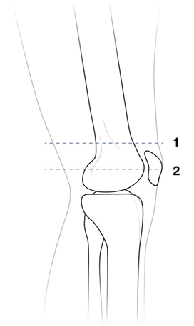
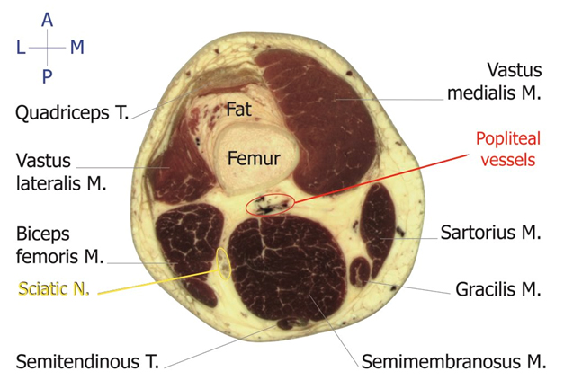
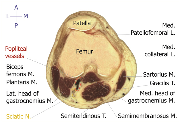
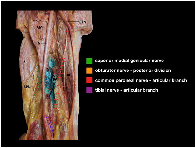
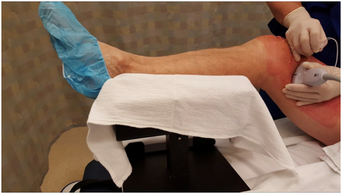
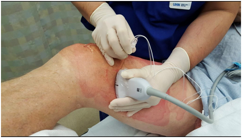
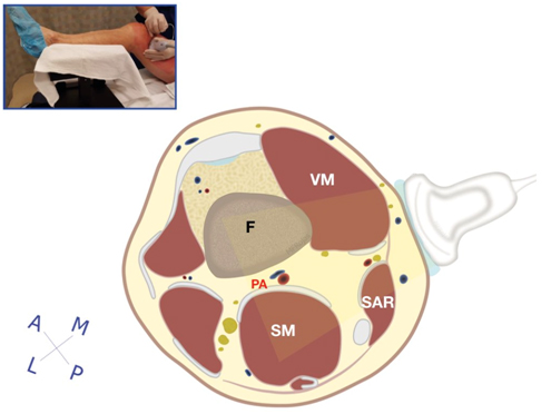
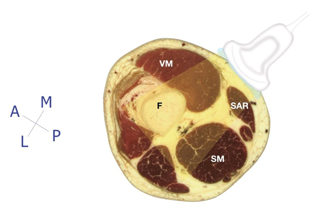
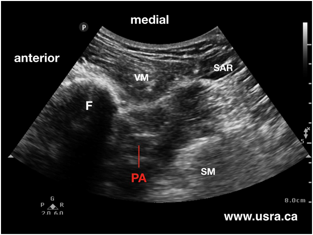
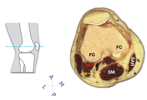
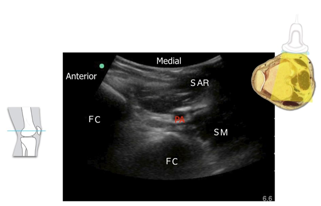
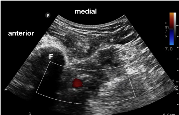
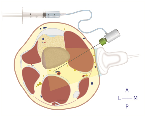
Background

The concept of local infiltration analgesia (LIA) was first introduced by Kohan and Kerr to provide pain relief following total knee arthroplasty (TKA). Today, this simple, surgeon performed peri-articular injection under direct vision has become commonplace in clinical practice around the world. Up to 150 mL of local anesthetic solution (often ropivacaine 0.2%) containing a variety of adjuncts (epinephrine, NSAID, opioid, steroid) is injected during and at the end of TKA. The injection targets the anterior and posterior capsule of the knee, intercondylar area, collateral ligaments, tissue along the femur and tibia and the subcutaneous tissue.

The analgesic effectiveness of the LIA technique is undisputed and has been confirmed in multiple clinical studies. The only drawback of single shot LIA is limited duration of analgesia (hours) unless liposomal bupivacaine is injected which can extend analgesia to up to 72 hours. Indwelling LIA catheter is not commonly applied because of the fear of infection.

The iPACK block was first introduced by Dr. Sanjay Sinha. The acronym iPACK stands for infiltration between popliteal artery and capsule of the knee. The block is performed by anesthesiologists under ultrasound guidance to provide pain relief to the posterior aspect of the knee after TKA by blocking the articular branches of the tibial, common peroneal and obturator nerves in the popliteal region.

The iPACK block likely achieves a similar degree of analgesia as posterior capsule injection as part of LIA performed by the surgeon. However, the iPACK block has a potential technical advantage over LIA injection since it is ultrasound guided and the needle path aims to stay clear of the popliteal artery and the sciatic nerve. In contrast, surgeons are often reluctant to inject deep into the posterior capsule for the fear of accidental popliteal artery puncture and sciatic nerve block since these posterior anatomical structures are not visualized anterior to the posterior capsule.

At the time of writing, there is no randomized controlled trial to compare anesthesiologist performed iPACK with surgeon performed posterior capsule LIA with respect to analgesic effectiveness and duration.

Anatomy

The iPACK block may be performed in the popliteal fossa about 1 finger breath above the patella (Figure 1, level 1) or more caudad at the level of the femoral condyle (Figure 1, level 2).

Figure 1. Levels of iPACK injection.
1 = 1 finger breath above the patella.
2 = level of the femoral condyles.

At the popliteal level above the patella (level 1 in Figure 1), a cross section of the thigh shows the following structures (Figure 2).

Figure 2. Cross sectional view of the thigh just above the patella.

More caudad in the popliteal fossa over the femoral condyle, a cross section of the thigh shows the following structures (Figure 3).

Figure 3. Cross sectional view of the thigh at the level of the femoral condyles.

The iPACK block aims to anesthetize the articular branches to the posterior aspect of the knee in the popliteal region (Figure 4).

Figure 4. Anatomical dissection showing articular branches of the tibial nerve, common peroneal nerve and obturator nerve to the posterior aspect of the knee.

Scanning Technique

  • Position the patient supine with the operative leg supported as shown below (Figure 5) and ultrasound position (Figure 6).
Figure 5. Leg support position for iPACK block.
  • After skin and transducer preparation, a curved 2-5 MHz transducer is applied transverse on the medial thigh approximately 1 to 2 finger breaths above the patella (Figure 6).
Figure 6. Transducer position over the medial aspect of the thigh approximately 1 finger breath above the patella.
  • The goal is to capture the best possible transverse and oblique view of the femoral shaft, the popliteal artery and the space posterior to the femoral shaft (Figure 7).
Figure 7. Schematic diagram showing transducer position and scanned area.
F = femoral shaft;
PA = popliteal artery;
SAR = sartorius muscle;
SM = semi-membranosus muscle;
VM = vastas medialis muscle
  • Optimize machine imaging capability by selecting the appropriate depth of field (usually within 7-8 cm), focus range and gain.
  • Visualize the outline of the femoral shaft as a curved hyper echoic line.
  • Also identify the pulsatile popliteal artery usually deep to the femoral shaft outline and a short distance posterior to the femur (Figure 8).
Figure 8. Sonogram of popliteal region above the patella.
F = femoral shaft;
PA = popliteal artery

Anatomical Correlation

Cross sectional view of the thigh approximately 1-2 finger breaths above the patella, anatomical dissection vs. ultrasound (Figures 9 and 10).

Figure 9. Cross section of the thigh showing anatomical structures immediately above the patella.
F = femur;
SAR = sartorius muscle;
SM = semi-membranosus muscle;
VM = vastus medialis muscle
Figure 10. Sonogram showing anatomical structures in the popliteal fossa at the same corresponding level.
F = femur;
PA = popliteal artery;
SAR = sartorius muscle;
SM = semi-membranosus muscle;
VM = vastus medialis muscle

Cross sectional view of the thigh at the level of the femoral condyle, anatomical dissection vs. ultrasound (Figures 11 and 12).

Figure 11. Cross section of the thigh showing anatomical structures at the level of the femoral condyle.
FC = femoral condyle;
PA = popliteal artery;
SAR = sartorius muscle;
SM = semi-membranosus muscle;
SN = sciatic nerve
Figure 12. Sonogram showing anatomical structures in the popliteal fossa at the level of the femoral condyle.
FC = femoral condyle;
PA = popliteal artery;
SAR = sartorius muscle;
SM = semi-membranosus muscle

Nerve Localization

  • Perform a systematic anatomical survey from anterior to posterior and superficial to deep. The posterior capsule of the knee is located between the femoral shaft and the popliteal artery.
  • With the transducer over the medial thigh (technique of Dr. Sanjay Sinha), identify the femoral shaft and the popliteal artery. The popliteal vein is often not seen. Note how far the artery is from the posterior aspect of the femur. This is the iPACK block approach at level 1 (1-2 finger breaths above the patella) (refer to Figure 1).
  • Use Colour Doppler to localize the popliteal artery (Figure 13)
Figure 13. Sonogram with Colour Doppler to visualize the popliteal artery.
F = femoral shaft;
Red area = popliteal artery
  • Move the transducer slightly more posterior to obtain a view of the femoral shaft near the edge of the screen. This slight scanning adjustment aims to identify a clear path for needle advancement. The goal is to avoid needle contact with the femoral shaft and unintentional popliteal artery puncture. Note that the desired needle insertion angle is often very steep (> 60o).
  • Note that the tibial and common peroneal nerves are not usually seen.

Needle Insertion Approach

  • Ultrasound guided iPACK block is considered an BASIC skill level block because this is a plane block.
  • The In Plane (IP) approach is recommended to visualize the needle shaft and tip movement during needle advancement.

In plane approach antero-medial to postero-lateral approach at level 1 (1 - 2 finger breaths above the patella) according to the technique of Dr. Sinha.

  • After skin and transducer preparation, a 8 cm 22 G echogenic block needle or a regular 22 G spinal needle is inserted into the medial thigh approximately 1 finger breath above the patella (Figure 14). Advance the needle often > 60 degree angle from the anterior end of the ultrasound transducer along its long axis in the postero-lateral direction.
Figure 14. Picture showing leg, transducer and needle insertion positions.
  • Observe real time needle advancement posterior to the femur and stay clear of the popliteal artery.
  • Aim to keep the needle immediately next to the femoral shaft at the start of needle advancement (Figure 15). The needle will invariably move posteriorly and laterally towards the popliteal artery as it is advanced deeper. Needle trajectory planning prior to actual needle insertion is therefore paramount.
  • Stop needle advancement once the needle tip has reached the lateral wall of the popliteal artery.
Figure 15. A schematic diagram showing the path and depth of needle advancement to reach the popliteal vessels.

Out of Plane Approach

An out of plane approach is possible but clinical experience with this approach has not been reported.

Local Anesthetic Injection

  • The goal is to deposit local anesthetic posterior to the femoral shaft in the interspace between the posterior capsule of the knee and the popliteal artery (Video 1).
  • Note that injection too deep i.e., beyond the popliteal vessels may result in anesthetic spillage to branches of the sciatic nerve affecting the common peroneal nerve more commonly than the tibial nerve in association with the needle insertion approach described above.
  • Inject a total of 10-15 mL local anesthetic incrementally as the needle is withdrawn slowly.
  • The goal is to deposit a wall of local anesthetic in the posterior femoral interspace without affecting the sciatic nerve and its branches.
  • In our centre, bupivacaine 0.25% is the anesthetic of choice but the optimal dose, concentration and volume of local anesthetic for iPACK block has not been determined. Neither has the optimal level of injection (above patella vs. at the femoral condyle level) been determined.

Clinical Pearls

Image Gallery

Video Gallery


Selected References

  • Kerr DR, Kohan L. Local infiltration analgesia: a technique for the control of acute postoperative pain following knee and hip surgery: a case study of 325 patients. Acta Orthop 2008;79(2):174-83.
  • Reddy AVG, Jangale A, Reddy RC, Sagi M, Gaikwad A, Reddy A. To compare effect of combined block of adductor canal block (ACB) with IPACK (Interspace between the Popliteal Artery and the Capsule of the posterior Knee) and adductor canal block (ACB) alone on Total knee replacement in immediate postoperative rehabilitation. International Journal of Orthopaedics Sciences 2017;3 (2c):141-145.
  • Sankineani, S.R., Reddy, A.R.C., Eachempati, K.K. et al. Comparison of adductor canal block and IPACK block (interspace between the popliteal artery and the capsule of the posterior knee) with adductor canal block alone after total knee arthroplasty: a prospective control trial on pain and knee function in immediate postoperative period. Eur J Orthop Surg Traumatol 2018.. https://doi.org/10.1007/s00590-018-2218-7
  • Thobhani S, Scalercio L, Elliott CE, Nossaman BD, Thomas LC, Yuratich D, Bland K, Osteen K, Patterson ME. Novel Regional Techniques for Total Knee Arthroplasty Promote Reduced Hospital Length of Stay: An Analysis of 106 Patients. Ochsner J 2017;17 (3):233-238.

Share to Facebook Share to Twitter More...