Youtube

Advancing the Science of Ultrasound Guided Regional Anesthesia and Pain Medicine

Catheter Technique

Performing A Sterile Procedure for Catheter Insertion


1. Sterilize the skin surface thoroughly with an antiseptic solution (e.g., 2% chlorhexidine in 70% alcohol).

2. Surround the block location with sterile drapes.

3. Place the transducer inside a sterile sheath.

4. Cover the transducer and cord completely inside the sterile sheath.

5. Hold the sheath in place with a rubber band around the transducer head.

6. Place the transducer over the sterile procedure field.

7. Use a package of sterile acoustic gel.

8. Apply sufficient amount of sterile gel over the site of scanning.

9. Place the transducer over the procedure site.

10. Insert a needle under ultrasound guidance.

Continuous Catheter Placement

  • There are two common approaches for catheter placement: the in plane and out of plane approaches. It is the author's preference to use the OUT OF PLANE needle approach for catheter insertion since the catheter is ideally positioned for advancement as it exits the tip of the needle parallel to the long axis of the target nerve.
  • Insertion using the IN PLANE needle approach is possible. However, this approach assumes that the catheter can turn 90 degrees upon exiting the tip of the needle to be advanced along the long axis of the nerve.
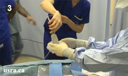
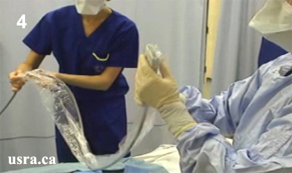
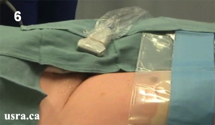
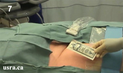
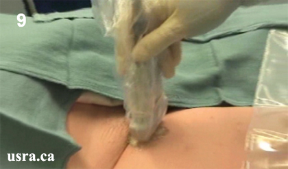
  • The ultrasound transducer and cord are covered completely inside a sterile sheath (see Preparing the Transducer for Single Shot).
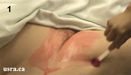
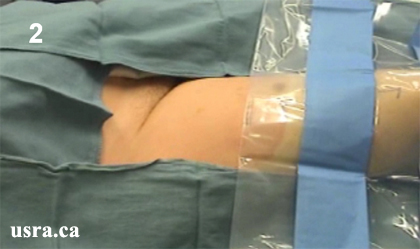
  • The technique of continuous catheter placement follows the same principle as the single shot injection technique. That is, the procedures for patient positioning, skin preparation and sterilization, and transducer selection are identical.
  • In general, a 17 G insulated needle is inserted perpendicular to the ultrasound transducer (Out of Plane Approach) and advanced to contact the target nerve.
  • Once the needle is deemed in contact with the target nerve (as indicated by nerve movement and/or nerve stimulation), inject 5-10 mL of local anesthetic or D5W (if nerve stimulation is desired) to distend the perineural space.
  • Distention of the perineural space will facilitate catheter threading especially in tight spaces e.g., interscalene groove.
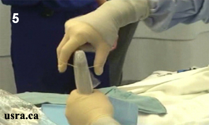
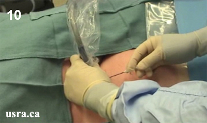
  • A 20 G stimulating or non stimulating catheter is then inserted 3-5 cm into the perineural space.
  • The catheter is often inserted without real time ultrasound guidance unless an assistant is available to hold the ultrasound transducer in place while the principal operator uses one hand to hold the needle and one hand to thread in the catheter.
  • It is often difficult to visualize the transverse view of the catheter which appears as a hyperechoic dot.
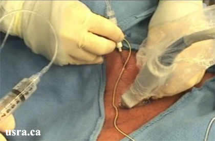
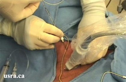
  • After the needle is withdrawn, real time assessment of local anesthetic spread is recommended during injection. Circumferential spread indicates that the catheter tip is located in an optimal position.
  • Suboptimal catheter position may be corrected by withdrawing the catheter a short distance before further local anesthetic is injected.
  • In the author's opinion, it is exceptionally challenging for the operator to purposefully advance the catheter under real-time ultrasound guidance. However, ultrasound is useful for differentiating circumferential from asymmetrical spread of local anesthetic injected through the catheter.

Video Presentation

How to Maximize Ultrasound Guidance for Catheter Placement

Sugantha Ganapathy, MBBS, DA, FRCA, FFARCS (I), FRCPC

Professor of Anesthesia
Director, Regional and Pain Research
University of Western Ontario
London, Ontario, Canada

Perineural Catheter Placement and Infusion Strategies

Geert J van Geffen, MD, PhD

Radboud University Medical Centre
Nijmegen, Netherlands

More videos available at our YouTube Channel - Click Here


Share to Facebook Share to Twitter More...