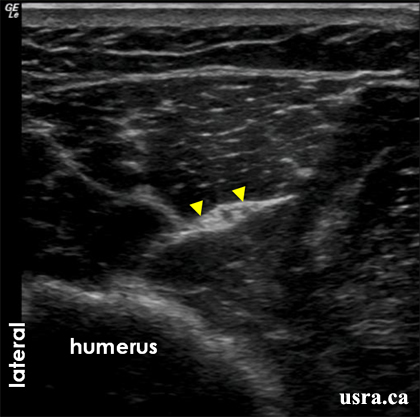
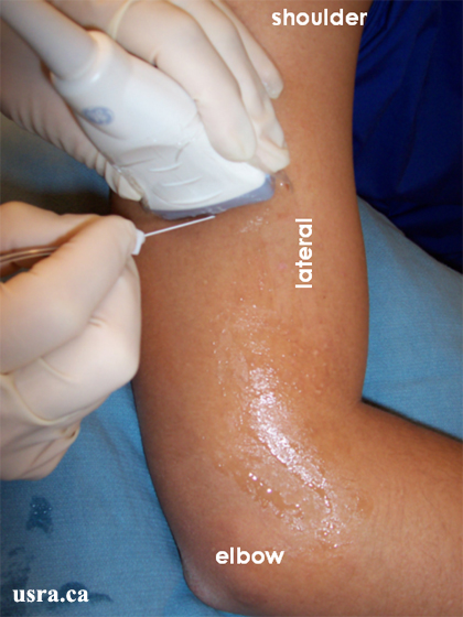

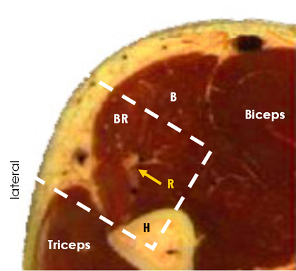
Radial Nerve Above the Elbow (4-5 cm Above)
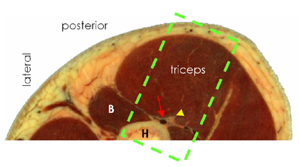
Anatomical Correlation
|
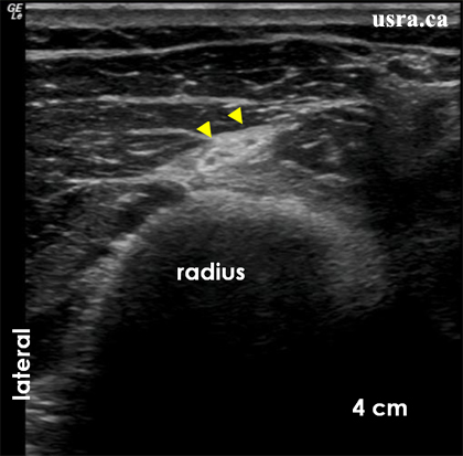
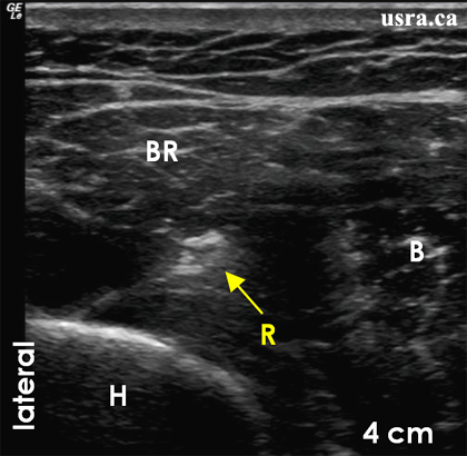
The solitary radial nerve appears predominantly hyperechoic at this region. B = brachialis muscle BR = brachioradialis muscle H = humerus R = radial nerve |
|
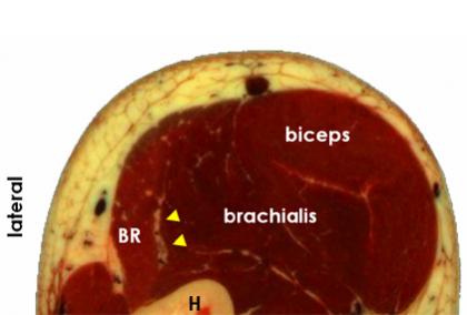
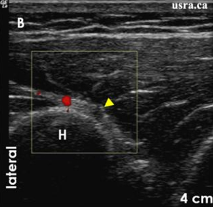
B = brachialis muscle BR = brachioradialis muscle Box = scan area H = humerus R = radial nerve |