Youtube

Advancing the Science of Ultrasound Guided Regional Anesthesia and Pain Medicine

Suprascapular Nerve Block

Introduction

The suprascapular nerve provides sensory innervation to the glenohumeral joint (shoulder). Suprascapular nerve block is indicated for relief of acute shoulder pain e.g., after shoulder surgery and is more effective when combined with blockade of the axillary nerve. It is also useful for the diagnosis and treatment of chronic shoulder pain secondary to bursitis, arthritis, degenerative joint and rotator cuff disease. Some studies show that blockade using local anesthetic and steroid can alleviate pain and disability in certain chronic shoulder pain conditions.

Anatomy

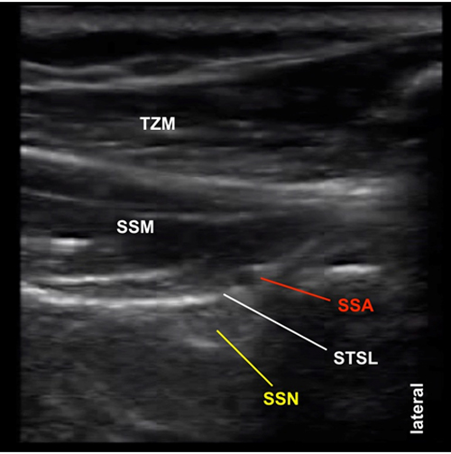
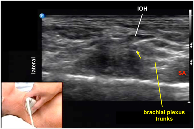
The suprascapular nerve (SSN) is a mixed nerve containing both motor and sensory fibers originating from the superior trunk of the brachial plexus (C5 and C6 nerve roots). Often it receives contribution from the C4 nerve root as well. The SSN passes underneath the omohyoid muscle in the posterior triangle of the neck. Then it passes posteriorly towards the scapula together with the omohyoid muscle towards the suprascapular notch. The nerve passes deep to the superior transverse scapular ligament through the scapular foramen into the supraspinous fossa. At the notch, the nerve is next to the suprascapular artery and vein but the vessels pass above the ligament (Figure 1 and 2).

Figure 1. A schematic diagram showing posterior view of the suprascapular nerve and vessels at the suprascapular notch

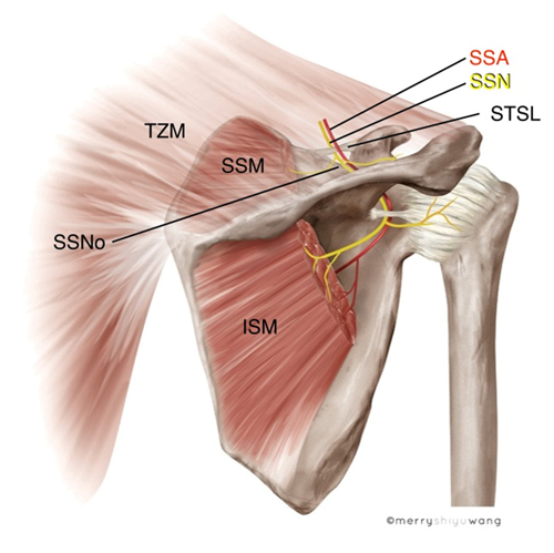
ISM = infraspinatus muscle
SSM = supraspinatus muscle
SSA = suprascapular artery
SSN = suprascapular nerve
TZM = trapezius muscle

Figure 2. Posterior view of the suprascapular nerve and surrounding structures at the suprascapular notch (cadaver dissection)

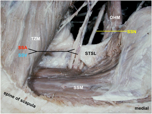
OHM = omohyoid muscle
SSM = supraspinatus muscle
SSA = suprascapular artery
SSV = suprascapular vein
SSN = suprascapular nerve
TZM = trapezius muscle
STSL = superior transverse scapular ligament

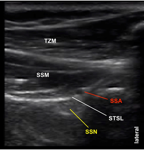
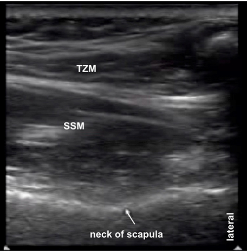
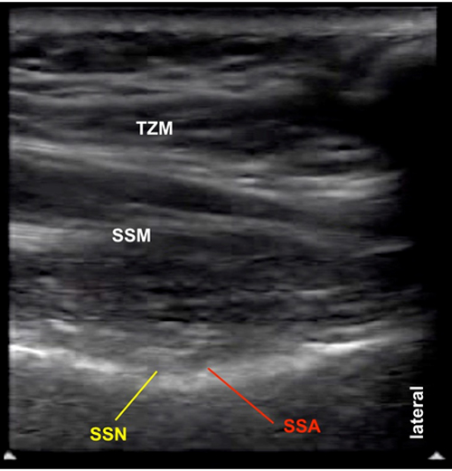
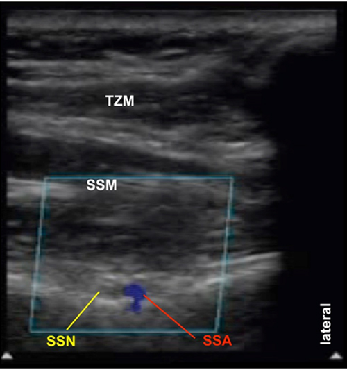
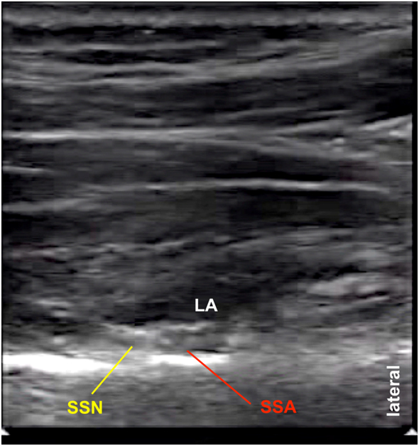
After traveling through the supraspinous fossa, the SSN reaches the spinoglenoid notch laterally and exits into the infraspinous fossa (Figure 3). Between the two notches, it is in direct contact with bone, covered by the inferior fascia of the supraspinatus muscle and accompanied by the suprascapular artery. The course of the SSN within the fossa is considerably oblique which has important impact on correct transducer positioning.

Figure 3. A schematic diagram showing superior view of the supraspinous fossa showing the course of the suprascapular nerve from the suprascapular notch to the spinoglenoid notch laterally

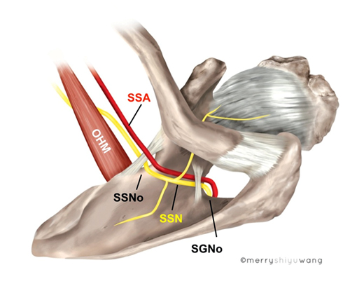
OHM = omohyoid muscle
SGNo = spinoglenoid notch
SSNo = suprascapular notch
SSA = suprascapular artery
SSN = suprascapular nerve

The suprascapular nerve innervates the supraspinatus and infraspinatus muscles. It also provides sensory innervation to both the glenohumeral and acromioclavicular joints, the coracohumeral ligament, the subacromial bursa and the scapula.

From an anatomical perspective, it is best to block the nerve over the supraspinous fossa half way between the suprascapular notch and the spinoglenoid notch.

Movie 2.1. Suprascapular nerve anatomy

Sonoanatomy

Sonographically, the suprascapular nerve often appears hyperechoic. At the suprascapular notch, the suprascapular nerve is located deep to the supraspinatus muscle and also deep to the suprascapular artery (SSA) and the superior transverse scapular ligament (STSL) as shown in Figure 4.

Note that this is NOT the site for SSN injection because the pleura is anterior to the suprascapular notch.

Figure 4. Sonoanatomy of suprascapular nerve and its surrounding structures over the suprascapular notch

SSM = supraspinatus muscle
SSA = suprascapular artery
SSN = suprascapular nerve
TZM = trapezius muscle
STSL = superior transverse scapular ligament

Scanning Technique

Position the patient sitting.

After skin and transducer preparation, place a linear 38-mm high frequency 10-12 MHz transducer on the scapula to obtain a best possible transverse view of the suprascapular nerve (SSN) and suprascapular vessels.

Optimize machine imaging capability by selecting the appropriate depth of field (usually within 3-5 cm), focus range and gain.

Ask the patient to place the hand over to the contralateral shoulder. This will move the scapula laterally to provide more space for SSN scanning. Also, this will move the target SSN injection site more laterally away from the thorax.

Position the transducer oblique (not parallel) to the spine of the scapula in the supraspinous fossa because the SSN runs an oblique course between the suprascapular notch and the spinoglenoid notch (Figure 5).

Place one end of the transducer over the scapular spine and the other end directing towards the coracoid process (Figure 5).

Figure 5. Ideal oblique transducer position with one end over the scapular spine and the other one pointing towards the coracoid process


The optimal transducer position is to scan the SSN at approximately 90 degrees to the course of the SSN from the suprascapular notch to the spinoglenoid notch (Figure 6). The finger under the transducer in Figure 6 imitates the direction in which the SSN runs. The transducer aims to capture a transverse view of the nerve.

Figure 6. Transducer position in relation to the simulated suprascapular nerve course (finger)


Nerve Localization

The external landmarks for transducer position and for localizing the SSN are: 1) the spine of the scapula; 2) the acromion and the acromial end of the clavicle and 3) the coracoid process.

There are 2 muscular and 3 bony internal sonographic landmarks. They are the trapezius muscle (TZ, more superficial) and the supraspinatus muscle (SSM). The bony landmarks are the supraspinous fossa, the suprascapular notch located anterior at the lateral part of the superior margin of the scapula) and the spinoglenoid notch located more postero-laterally.

First identify the SSM, which is thicker than the overlying trapezius muscle in the supraspinous fossa (Figure 7).

The floor of the supraspinous fossa is visualized as a continuous hyperechoic line deep to the SSM (Figure 7).

Figure 7. Sonogram showing the trapezius and supraspinatus muscles

SSM = supraspinatus muscle
TZM = trapezius muscle

Then move the transducer laterally to visualize the acromial end of the clavicle and/or the acromion; both cast a superficial bony shadow (Figure 8). Note that the trapezius muscle attaches to the acromion.

Figure 8. Sonogram showing the acromion when the transducer is moved more laterally

AC = acromion
SSM = supraspinatus muscle
TZM = trapezius muscle

Slowly turn the outer end of the transducer towards the tip of the palpable coracoid process anteriorly. This is often the optimal transducer position to visualize the SSN. The transducer is somewhat oblique to the scapular spine and not parallel to the spine.

Then slowly angle the transducer anteriorly to visualize the suprascapular notch. Sonographically, this bony landmark is represented by a break in the supraspinous fossa line. At the suprascapular notch region, one can visualize the SSN within the scapular foramen (Figure 9). The suprascapular artery runs above the SSN and the superior transverse scapular ligament; however, the STSL is not always visualized under ultrasound. Note that this location is NOT the optimal site for SSN block because the pleura is located anterior to the suprascapular notch. Accidental anterior advancement of the needle can possibly puncture the pleura causing pneumothorax.

Figure 9. Sonogram showing the suprascapular nerve in the suprascapular notch

SSM = supraspinatus muscle
SSA = suprascapular artery
SSN = suprascapular nerve
TZM = trapezius muscle
STSL = superior transverse scapular ligament

Then follow the supraspinous fossa slightly posteriorly to find the neck of the scapula which shows up as a depression of the supraspinous fossa bone shadow (Figure 10).

Figure 10. Sonogram showing the neck of the scapula in the supraspinous fossa

SSM = supraspinatus muscle
TZM = trapezius muscle

The neck of the scapula will lead to the spinoglenoid notch when scan more laterally and posteriorly (Figure 11). The notch here appears as a deeper depression of the supraspinous fossa bony outline.

Figure 11. Sonogram showing the spinoglenoid notch as a deep depression in the supraspinous fossa

SSM = supraspinatus muscle

The optimal location to perform SSN block is half way between the scapular notch and the spinoglenoid notch. At this location, the suprascapular notch is not seen. The SSN is located underneath the deep fascia of the SSM and next to the suprascapular artery (Figure 12). The suprascapular artery is no longer above the SSN at this location.

Figure 12. Sonogram showing the suprascapular nerve next to the suprascapular artery in the supraspinous fossa

SSM = supraspinatus muscle
SSA = suprascapular artery
SSN = suprascapular nerve
TZM = trapezius muscle

At the optimal block location, the SSN lies next to the suprascapular artery (Figure 12). The suprascapular artery is identified using Color Doppler (Figure 13).

Figure 13. Color Doppler showing the suprascapular artery lateral to the suprascapular nerve

SSM = supraspinatus muscle
SSA = suprascapular artery
SSN = suprascapular nerve
TZM = trapezius muscle

Needle Insertion Approach
Ultrasound guided suprascapular nerve block is considered an intermediate skill level block.

The goal is to block the SSN approximately halfway in between the suprascapular and the spinoglenoid notches. There is no need to visualize the notches at the time of injection.

We recommend an in-plane needle approach aiming from posteromedial to anterolateral. Needle insertion from the lateral side, on the other hand, is more challenging because the acromion is in the path of the needle making it necessary to insert the needle at a steep angle thus reduces needle visibility (Figure 14).

Figure 14. In plane needle insertion approach in the postero-medial to anterolateral direction


With the patient sitting up, a 22 G, 8 cm needle is advanced to penetrate the trapezius and supraspinatus muscles until the needle is positioned immediately next to the SSN and SSA (Figure 15).

Figure 15. Sonogram showing the needle tip in the supraspinatus muscle heading towards the suprascapular nerve

arrow = needle tip

With either needle approach, it is important to hydrodissect repeatedly during needle advancement to rule out injection into the SSM (intramuscular injection). The optimal needle endpoint is reached when the needle tip is positioned underneath the fascia of the SSM.

Avoid targeting the SSN in the suprascapular notch because accidental anterior needle advancement can puncture the pleura anteriorly.

Movie 2.2. In plane needle advancement towards the suprascapular nerve

Local Anesthetic Injection

The goal is to first inject local anesthetic deep to the fascia of the SSM. A total of 5-8 mL of local anesthetic is usually sufficient to block the SSN (Figure 16).

If local anesthetic spread is detected initially within the SSM, it is necessary to advance the needle further to traverse the fascia of the SSM.

Figure 16. Sonogram showing a local anesthetic bolus injected immediately above the suprascapular nerve and artery

LA = local anesthetic
SSA = suprascapular artery
SSN = suprascapular nerve

Movie 2.3. In plane needle insertion and local anesthetic injection

Clinical Pearls

Blocking the Suprascapular Nerve in the Posterior Cervical Triangle (an Alternative Approach)

It is possible to block the SSN more centrally in the posterior cervical triangle after the nerve has branched off the superior trunk of the brachial plexus.

When the transducer is positioned in the supraclavicular fossa, the SSN can be visualized as a round to oval hypoechoic structure deep to the inferior belly of the omohyoid muscle (a reliable internal sonographic landmark) and lateral to the superior trunk (Figure 17). Note that the appearance of the SSN is quite different at this location compared to the supraspinous fossa.

Figure 17. Sonogram showing the suprascapular nerve branching off from the superior trunk of the brachial plexus

arrow = suprascapular nerve
IOH = inferior belly of omohyoid muscle
SA = subclavian artery

For SSN block at this location, the nerve is traced more postero-laterally before injection at a considerable distance from the superior trunk of the brachial plexus. This will avoid unintentional blockade of the superior trunk. It is also advisable to limit local anesthetic injection to a smaller volume e.g., 2 mL.

Note that there may be another hypoechoic structure next to the SSN. This is either the superficial cervical artery (SCA), or the suprascapular artery which is more posteriorly and inferiorly (Figure 18). The vessels must be differentiated from the SSN using Color Doppler.

Figure 18. Sonogram showing the superficial cervical artery immediately next to the suprascapular nerve

arrow = suprascapular nerve

Video Gallery

Selected References

  • Battaglia PJ, Haun DW, Dooley K, Kettner NW. Sonographic measurement of the normal suprascapular nerve and omohyoid muscle. Man Ther 2014;19:165-8.
  • Borglum J, Bartholdy A, Hautopp H, Krogsgaard MR, Jensen K. Ultrasound-guided continuous suprascapular nerve block for adhesive capsulitis: one case and a short topical review. Acta Anaesthesiol Scand 2011;55:242-7.
  • Chan C-W, Peng PWH. Suprascapular nerve block: a narrative review. Reg Anesth Pain Med 2011;36:358-73.
  • Draeger RW, Messer TM. Suprascapular nerve palsy following supraclavicular block for upper extremity surgery: report of 3 cases. J Hand Surg Am 2012;37:2576-9.
  • Elsharkawy HA, Abd-Elsayed AA, Cummings KC, Soliman LM. Analgesic efficacy and technique of ultrasound-guided suprascapular nerve catheters after shoulder arthroscopy. Ochsner J 2014;14:259-63.
  • Hackworth RJ. A new and simplified approach to target the suprascapular nerve with ultrasound. J Clin Anesth 2013;25:347-8.
  • Herring AA, Stone MB, Nagdev A. Ultrasound-guided suprascapular nerve block for shoulder reduction and adhesive capsulitis in the ED. Am J Emerg Med 2010.
  • Ko SH, Kang BS, Hwang CH. Ultrasonography- or electrophysiology-guided suprascapular nerve block in arthroscopic acromioplasty: a prospective, double-blind, parallel-group, randomized controlled study of efficacy. Arthroscopy 2013;29:794-801.
  • Peng PWH, Wiley MJ, Liang J, Bellingham GA. Ultrasound-guided suprascapular nerve block: a correlation with fluoroscopic and cadaveric findings. Can J Anaesth 2010;57:143-8.
  • Siegenthaler A, Moriggl B, Mlekusch S, Schliessbach J, Haug M, Curatolo M, Eichenberger U. Ultrasound-Guided Suprascapular Nerve Block, Description of a Novel Supraclavicular Approach. Reg Anesth Pain Med 2012.
  • Soneji N, Peng PWH. Ultrasound-guided pain interventions - a review of techniques for peripheral nerves. Korean J Pain 2013;26:111-24.
  • Taskaynatan MA, Ozgul A, Aydemir K, Koroglu OO, Tan AK. Accuracy of ultrasound-guided suprascapular nerve block measured with neurostimulation. Rheumatol Int 2011.
  • Yucesoy C, Akkaya T, Ozel O, Comert A, Tüccar E, Bedirli N, Unlu E, Hekimoglu B, Gumus H. Ultrasonographic evaluation and morphometric measurements of the suprascapular notch. Surg Radiol Anat 2009;31:409-14.

Share to Facebook Share to Twitter More...