Youtube

Advancing the Science of Ultrasound Guided Regional Anesthesia and Pain Medicine

Lateral Femoral Cutaneous Nerve Block

Anatomy

The lateral femoral cutaneous nerve (LFCN) arises from the dorsal divisions of L2-3. After emerging from the lateral border of the psoas major muscle, it courses inferiorly and laterally towards the anterior superior iliac spine (ASIS). It then passes under the inguinal ligament and over the sartorius muscle into the thigh, where it divides into two branches (anterior and posterior). The LFCN, a small subcutaneous nerve located between the fascia lata and iliaca, provides sensory innervation to the lateral thigh.

Scanning Technique

  • Position the patient supine with the leg extended in the neutral position.
  • Expose the groin and mark the anterior superior iliac spine (ASIS).
  • After skin and transducer preparation, place a linear transducer with the appropriate frequency range (10-12 MHz) immediately medial to the ASIS along the inguinal ligament. The lateral end of the transducer is placed on the ASIS. Scan medially and inferiorly from the ASIS.
  • Optimize machine imaging capability; select appropriate depth of field (usually within 1-2 cm), focus range and gain.
Transducer medial and inferior to the right anterior superior iliac spine (ASIS) and inferior to the inguinal ligament (IL).

Anatomical Correlation

Coming Soon

Nerve Localization

  • Perform a systematic anatomical survey medial and inferior to the ASIS. The LFCN is expected to lie in the subcutaneous plane deep to the fascia lata below the ASIS.
  • Identify the ASIS which casts a bony shadow. Then move the transducer medially and inferiorly to locate the fascia lata and the sartorius muscle. The LFCN, a small hypoechoic structure is often found in the interfascial plane above the sartorius muscle as shown in figure below.
Sonogram of the right lateral femoral cutaneous nerve (arrowhead) below the inguinal crease.

SAR = sartorius muscle
FL = facia lata
  • Percutaneous nerve stimulation further confirms nerve identity.
  • Another good location to visualize the LFCN is approximately 3 fingerbreadths inferior (caudad) to the ASIS. At this distal location, the nerve is often visualized in a hypoechoic fat filled subfascial space between the sartorius muscle medially and the tensor fascia lata muscle laterally. The nerve may be then traced proximally i.e., cephalad as it gets smaller and smaller and changes from oval to linear until it reaches on top of the sartorius muscle.
Sonogram of lateral femoral cutaneous nerve (arrowhead) 3 finger breadths caudal to the ASIS
Sonogram of lateral femoral cutaneous nerve after division into anterior and posterior branches (arrowhead)

Needle Insertion Approach

In Plane Approach

  • Ultrasound guided LFCN block is considered an INTERMEDIATE skill level block because the nerve is small and may not be clearly visualized in the subcutaneous plane.
  • Insert a 5 cm 22 G needle inline with the transducer and the ultrasound beam.
  • Insert the needle at a shallow angle to enter the skin surface to reach the plane just below the fascia lata immediately medial and inferior to the ASIS.
  • If the nerve cannot be visualized, place the needle immediately medial and inferior to the ASIS close to where the sartorius muscle attaches to the ASIS.
  • Hydro dissection with saline in the plane below the fascia lata may enhance the visibility of LFCN after introduction of a hypoechoic fluid collection.
Pre Injection

* = lateral femoral cutaneous nerve
ASIS = anterior superior iliac spine
FL = fascial lata
SAR = sartorius muscle
Post Injection

* = lateral femoral cutaneous nerve
ASIS = anterior superior iliac spine
FL = fascial lata
SAR = sartorius muscle
Arrowheads = needle

A hypoechoic local anesthetic collection is now seen below FL.

Out of Plane Approach

Alternatively, an out of plane approach is just as simple to perform as the in plane approach.

Local Anesthetic Injection

  • In general, a small volume (e.g., 2-3 mL) local anesthetic injection is indicated for a diagnostic LFCN block.
  • It is important to note that this is a subfascial injection (below the fascia lata) and not a subcutaneous injection.

Clinical Pearls

Pitfalls in Nerve Localization

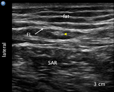
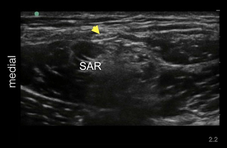
It is also common to visualize hyperechoic structures within the sartorius muscle. It is important not to mistake the hyperechoic tendinous part of the sartorius muscle for the LFCN. Differentiation is possible by performing a systematic dynamic scan to trace the nerve in the fat filled space below the ASIS from distal to proximal and vice versa.

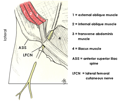
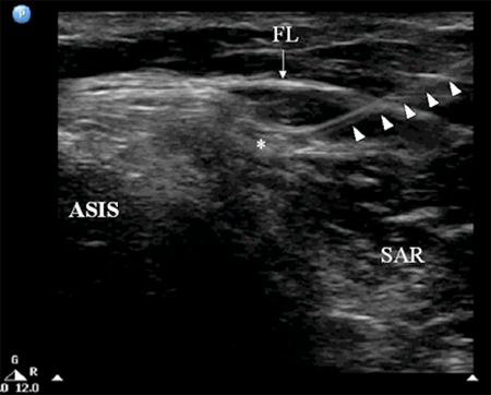
Course of the Lateral Femoral Cutaneous Nerve

The whole course of the LFCN as well as its formation is highly variable among individuals. Thus the LFCN may not be found immediately medial to the ASIS. Other practical important locations to locate the LFCN are:

  1. the nerve may be far more medially located, e.g., halfway between the ASIS and the pubic symphysis or tubercle; in this case, the LFCN is sandwiched between fascia lata and fascia iliaca before it comes on top of SM
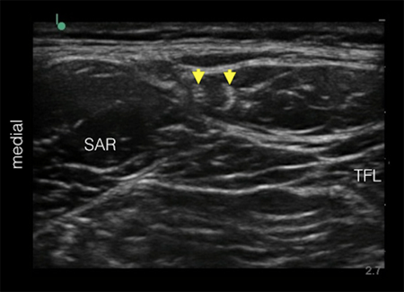
  2. the nerve may course over the iliac crest lateral to the ASIS;
  3. very rarely, the LFCN runs through or deep to the substance of the sartorius muscle (e.g., if the nerve is arising from the femoral nerve). In these cases, tendinous part of the sartorius muscle may be mistaken for the nerve.

Note that regardless of anatomical variations, the key to success for locating the LFCN is to first locate the nerve distally in the fat filled space and then trace the nerve proximally towards the ASIS.

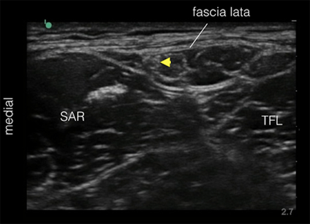
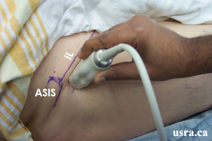
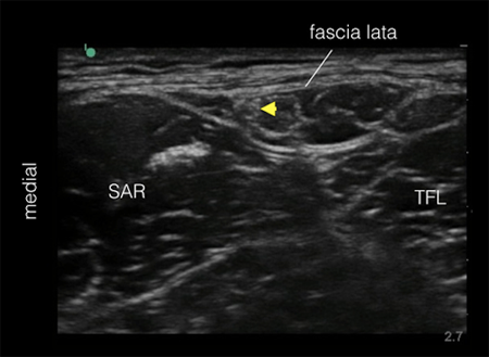
Nerve Located Between the Tensor Fascia Lata and Sartorius Muscles

The lateral femoral cutaneous nerve is usually visualized as a relatively hyperechoic nerve structure in the proximal thigh (several cm inferior to the anterior superior iliac spine) within a fat filled hypoechoic space. This space is found between the sartorius muscle medially and the tensor fascia lata muscle laterally. The nerve is covered by the fascia lata, thus the nerve is subfascial. Note that the nerve is not subcutaneous.

Catheter Insertion

Catheter insertion is not indicated for this block.

Image Gallery

Image of lateral femoral cutaneous nerve when scan proximally

Sonogram showing lateral femoral cutaneous nerve over the sartorius muscle when scan proximally

Image of lateral femoral cutaneous nerve when scan distally


A. Before branching

B. After branching

Video Gallery

Selected References

  • Moritz T, Prosch H, Berzaczy D, Happak W, Lieba-Samal D, Bernathova M, Auff E, Bodner G. Common anatomical variation in patients with idiopathic meralgia paresthetica: a high resolution ultrasound case-control study. Pain Physician 2013;16:E287-93.
  • Shteynberg A, Riina LH, Glickman LT, Meringolo JN, Simpson RL. Ultrasound guided lateral femoral cutaneous nerve (LFCN) block: safe and simple anesthesia for harvesting skin grafts. Burns 2013;39:146-9.
  • Zhu J, Zhao Y, Liu F, Huang Y, Shao J, Hu B. Ultrasound of the lateral femoral cutaneous nerve in asymptomatic adults. BMC Musculoskelet Disord 2012;13:227.
  • Aravindakannan T, Wilder-Smith EP. High-resolution ultrasonography in the assessment of meralgia paresthetica. Muscle Nerve. 2012;45:434-5.
  • Tagliafico A, Serafini G, Lacelli F, Perrone N, Valsania V, Martinoli C. Ultrasound-guided treatment of meralgia paresthetica (lateral femoral cutaneous neuropathy): technical description and results of treatment in 20 consecutive patients. J Ultrasound Med 2011;30:1341-6.
  • Kim JE, Lee SG, Kim EJ, Min BW, Ban JS, Lee JH. Ultrasound-guided Lateral Femoral Cutaneous Nerve Block in Meralgia Paresthetica. Korean J Pain 2011;24:115-8.
  • Hara K, Sakura S, Shido A. Ultrasound-guided lateral femoral cutaneous nerve block: comparison of two techniques. Anaesth Intensive Care 2011;39:69-72.
  • Bodner G, Bernathova M, Galiano K, Putz D, Martinoli C, Felfernig M. Ultrasound of the lateral femoral cutaneous nerve: normal findings in a cadaver and in volunteers. Reg Anesth Pain Med 2009;34:265-8.
  • Damarey B, Demondion X, Boutry N, Kim H-J, Wavreille G, Cotten A. Sonographic assessment of the lateral femoral cutaneous nerve. J Clin Ultrasound 2009;37:89-95.
  • Tumber PS, Bhatia A, Chan VW. Ultrasound-guided lateral femoral cutaneous nerve block for meralgia paresthetica. Anesth Analg 2008;106:1021-2.
  • Ng I, Vaghadia H, Choi PT, Helmy N. Ultrasound imaging accurately identifies the lateral femoral cutaneous nerve. Anesth Analg 2008;107:1070-4.
  • Hurdle MF, Weingarten TN, Crisostomo RA, Psimos C, Smith J. Ultrasound-guided blockade of the lateral femoral cutaneous nerve: technical description and review of 10 cases. Arch Phys Med Rehabil 2007;88:1362-4.

Share to Facebook Share to Twitter More...