Youtube

Advancing the Science of Ultrasound Guided Regional Anesthesia and Pain Medicine

Thoracic Paravertebral Block

General

Authors

Yasuyuki Shibata M.D.
Department of Anesthesiology
Nagoya University Hospital
Nagoya, Japan
Ki Jinn Chin FRCPC
Department of Anesthesia
University of Toronto
 

The resurgence of interest in thoracic paravertebral block (TPVB) can be traced back to a seminal article by Eason and Wyatt in 1979,1 although it was first described by Selheim in 1906 and modified by Lawen in 1911.2 The conventional technique of TPVB involves inserting the needle perpendicular to all planes, contacting the transverse process, and then walking off it with the needle. The commonly-used endpoints for needle insertion include loss-of-resistance to air or saline,1,2 advancing a pre-determined distance,3 or neurostimulation.4,5 Complications of TPVB are reportedly low, with the most feared being pleural puncture and pneumothorax (1.1% and 0.5% respectively in 1 study6).

Ultrasound has been used to enhance the safety and efficacy of TPVB by determining the location and depth of the transverse process and the parietal pleura.7,8 Pusch et al8 performed a pre-procedure scan and then performed TPVB in the conventional fashion using loss-of-resistance to saline as their endpoint. Hara et al7 imaged the transverse process and the thoracic paravertebral space (TPVS) in a longitudinal parasagittal plane during actual block performance; however the needle was inserted out-of-plane to the probe using a conventional technique, and loss-of-resistance to saline was used as the endpoint for needle placement rather than ultrasonographic visualization of needle-tip position.

Here we describe an in-plane ultrasound-guided technique that utilizes direct visualization of needle-tip position and local anesthetic spread as the endpoint. The approach is similar to one described by Kappis in 1912. In the original description, a 10 cm needle was inserted three fingerbreadths from the midline at a 45 degree angle to the skin, and advanced into the TPVS until the tip contacted the thoracic vertebral body. The technique was eventually abandoned because of the risk of needle penetration through the intervertebral foramen, and thus inadvertent dural puncture and spinal cord injury. However ultrasonographic visualization of in-plane needle advancement reduces the risk of pleural puncture as well as entry of the needle into the intervertebral foramen. Nevertheless the practitioner should be aware that there is a risk of epidural local anesthetic spread with paravertebral injection.

Anatomy

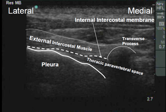
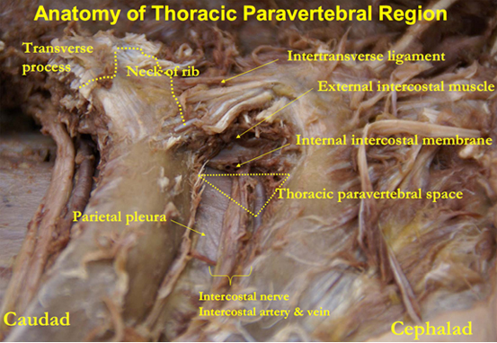
The thoracic paravertebral space (TPVS), when viewed in transverse cross-section is triangular-shaped (red triangle in figure below). The base is formed by the posterolateral aspect of the vertebral body / intervertebral discs / intervertebral foramina / articular processes. The anterolateral border is formed by the parietal pleura, whilst the posterior border is formed by the superior costotransverse ligament. This ligament extends from the inferior aspect of the transverse process above to the superior aspect of the rib tubercle below. Lateral to this ligament (and continuous with it) is the internal intercostal membrane, which is the aponeurotic continuation of the internal intercostal muscle, and thus runs between the upper and lower border of adjacent ribs.12 The apex of the triangular TPVS communicates with the intercostal space laterally. The cephalad limit of the TPVS has not been defined. It has been shown that solution injection into the TPVS can spread caudad into the abdominal and lumbar region, through the medial and lateral arcuate ligaments of the diaphragm. It is generally accepted, however, that the caudad limit of the paravertebral space is at the origin of the psoas muscle at L1.9


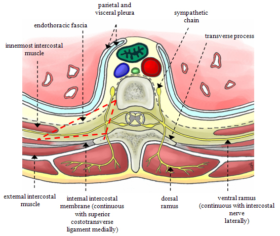
The TPVS contains mainly fatty tissue, and is traversed by the intercostal or spinal nerves, intercostal vessels, dorsal rami, rami communicantes, and the sympathetic chain. The spinal nerves do not have a fascial sheath in the TPVS, which explains their susceptibility to local anesthetic blockade.

The space is divided into an anterior and posterior compartment by a fibroelastic membrane, the endothoracic fascia. The endothoracic fascia is the deep investing fascia of the thoracic cavity. It blends medially with the periosteum of the vertebral body; and laterally, is closely applied to the ribs. Caudally, it is continuous with the transversalis fascia of the abdominal cavity and this may explain why solutions injected in the TPVS may spread to the lumbar region. The spinal nerves have been described as running through the compartment posterior to the endothoracic fascia.2 This however is controversial,10,11 as the precise anatomy of the endothoracic fascia, and its relationship to the spinal nerves in particular, remains ill-defined. It has been shown that injection closer to the spinal nerves (using a nerve-stimulator-guided technique) is more likely to result in longitudinal spread of the injectate in the TPVS.11

Scanning Technique

  • Place the patient in one of the following positions to expose their upper back: a) lateral decubitus position (surgical side uppermost);
    b) prone position;
    c) sitting position.
  • After skin and transducer preparation (see Preparing Transducer for Single Shot), place a linear 38 mm, high frequency 10-12 MHz transducer in an axial (transverse) plane on the rib at the selected thoracic level, just lateral to the spinous process.
 
  • Optimize machine imaging capability by selecting the appropriate depth of field (within 2-3 cm), focus range and gain.
  • The transverse process and rib are visualized as a hyperechoic line with acoustic shadowing below it (figure below).
 

Thoracic Paravertebral Space Localization

  • Move the transducer caudad into the intercostal space between adjacent ribs.
  • The transverse process can be visualized on the medial side as a hyperechoic convex line with acoustic shadowing beneath (figure below).
  • The TPVS and the adjoining intercostal space can now be visualized (figure below).
  • The TPVS is a wedge-shaped hypoechoic layer demarcated by the hyperechoic lines of the pleura below and the internal intercostal membrane above.
  • Note that the pleura is clearly visible as a hyperechoic line that moves with respiration and with underlying hyperechoic air artifacts. This is distinctly different from the hyperechoic rib line with underlying acoustic shadow.
  • It may be possible to identify intercostal vessels in the TPVS with Color Doppler, and if so, these should be avoided.

Anatomical Correlation

Coming Soon

Nerve Localization

Coming Soon

Needle Insertion Approach

  • Ultrasound guided thoracic paravertebral block is considered an INTERMEDIATE skill level block.
  • Accurate needle tip visualization is ESSENTIAL at all times to avoid inadvertent pleural puncture or entry into the intervertebral foramen.

In Plane Approach (Lateral to Medial)

  • Following local anesthetic infiltration of the skin, insert a 20-gauge Tuohy needle at the outer (lateral) end of the transducer, IN-PLANE with the ultrasound beam.
  • It is recommended that the bevel of the Tuohy needle tip be oriented upwards towards the transducer as this may reduce the risk of penetrating injury to intercostal vessels, nerve or pleura in the event of inadvertent needle contact.
 
  • Advance the needle in-plane with the transducer, in a lateral-to-medial direction.
  • Advance the needle (arrows, figure below) under real time ultrasound guidance and visualize needle tip penetration through the internal intercostal membrane and its entry into the TPVS.
  • A pop is often felt as the needle penetrates the internal intercostal membrane.
 

Out of Plane Approach

The out of plane approach is an acceptable approach for this block.

Local Anesthetic Injection

  • After ensuring negative aspiration for blood, 15-20 mL of local anesthetic is injected slowly in small increments into the TPVS.
  • The TPVS will be observed to distend, pushing the pleura downwards (ventrally) (figure below).

Clinical Pearls

Coming Soon

Catheter Insertion

Image Gallery

Coming Soon

Video Gallery

Selected References

  • Eason MJ, Wyatt R. Paravertebral thoracic block-a reappraisal. Anaesthesia 1979;34(7):638.
  • Karmakar MK. Thoracic paravertebral block. Anesthesiology 2001;95(3):771.
  • Greengrass R, O'Brien F, Lyerly K, et al. Paravertebral block for breast cancer surgery. Can J Anaesth 1996;43(8):858.
  • Lang SA. The use of a nerve stimulator for thoracic paravertebral block. Anesthesiology 2002;97(2):521; author reply 521.
  • Naja MZ, Ziade MF, Lonnqvist PA. Nerve-stimulator guided paravertebral blockade vs. general anaesthesia for breast surgery: a prospective randomized trial. Eur J Anaesthesiol 2003;20(11):897.
  • Lonnqvist PA, MacKenzie J, Soni AK, et al. Paravertebral blockade. Failure rate and complications. Anaesthesia 1995;50(9):813.
  • Hara K, Sakura S, Nomura T, et al. Ultrasound guided thoracic paravertebral block in breast surgery. Anaesthesia 2009;64(2):223.
  • Pusch F, Wildling E, Klimscha W, et al. Sonographic measurement of needle insertion depth in paravertebral blocks in women. Br J Anaesth 2000;85(6):841.
  • Lonnqvist PA, Hildingsson U. The caudal boundary of the thoracic paravertebral space. A study in human cadavers. Anaesthesia 1992;47(12):1051.
  • Lang SA, Saito T. Thoracic paravertebral nerve block, nerve stimulator guidance and the endothoracic fascia. Anaesthesia 2005;60(9):930.
  • Vallieres E. The costovertebral angle. Thorac Surg Clin 2007;17:503-510.
  • Naja MZ, Ziade MF, El Rajab M, et al. Varying anatomical injection points within the thoracic paravertebral space: effect on spread of solution and nerve blockade. Anaesthesia 2004;59(5):459.

Share to Facebook Share to Twitter More...